Anti-SUZ12 antibody (ab12073)
Key features and details
- Rabbit polyclonal to SUZ12
- Suitable for: WB
- Knockout validated
- Reacts with: Human
- Isotype: IgG
Overview
-
Product name
Anti-SUZ12 antibody
See all SUZ12 primary antibodies -
Description
Rabbit polyclonal to SUZ12 -
Host species
Rabbit -
Specificity
Antibody has been shown to recognise endogenous Suz12 in Colon cancer and Breast cancer cell line (See image below) -
Tested Applications & Species
See all applications and species dataApplication Species WB Human -
Immunogen
Synthetic peptide corresponding to Human SUZ12 aa 700 to the C-terminus conjugated to keyhole limpet haemocyanin.
(Peptide available asab12389)
Properties
-
Form
Liquid -
Storage instructions
Shipped at 4°C. Store at +4°C short term (1-2 weeks). Upon delivery aliquot. Store at -20°C or -80°C. Avoid freeze / thaw cycle. -
Storage buffer
pH: 7.40
Preservative: 0.02% Sodium azide
Constituent: PBS
Batches of this product that have a concentration Concentration information loading...
Purity
Immunogen affinity purifiedClonality
PolyclonalIsotype
IgGResearch areas
Associated products
-
ChIP Related Products
-
Compatible Secondaries
-
Isotype control
-
KO cell lines
-
KO cell lysates
Applications
The Abpromise guarantee
Our Abpromise guarantee covers the use of ab12073 in the following tested applications.
The application notes include recommended starting dilutions; optimal dilutions/concentrations should be determined by the end user.
GuaranteedTested applications are guaranteed to work and covered by our Abpromise guarantee.
PredictedPredicted to work for this combination of applications and species but not guaranteed.
IncompatibleDoes not work for this combination of applications and species.
Application Species WB HumanAll applications MouseCommon marmosetApplication Abreviews Notes WB (9) Use a concentration of 1 - 5 µg/ml. Detects a band of approximately 95 kDa (predicted molecular weight: 83 kDa).Notes WB
Use a concentration of 1 - 5 µg/ml. Detects a band of approximately 95 kDa (predicted molecular weight: 83 kDa).Target
-
Function
Polycomb group (PcG) protein. Component of the PRC2/EED-EZH2 complex, which methylates 'Lys-9' (H3K9me) and 'Lys-27' (H3K27me) of histone H3, leading to transcriptional repression of the affected target gene. The PRC2/EED-EZH2 complex may also serve as a recruiting platform for DNA methyltransferases, thereby linking two epigenetic repression systems. Genes repressed by the PRC2/EED-EZH2 complex include HOXC8, HOXA9, MYT1 and CDKN2A. -
Tissue specificity
Overexpressed in breast and colon cancer. -
Involvement in disease
Note=A chromosomal aberration involving SUZ12 may be a cause of endometrial stromal tumors. Translocation t(7;17)(p15;q21) with JAZF1. The translocation generates the JAZF1-SUZ12 oncogene consisting of the N-terminus part of JAZF1 and the C-terminus part of SUZ12. It is frequently found in all cases of endometrial stromal tumors, except in endometrial stromal sarcomas, where it is rarer. -
Sequence similarities
Belongs to the VEFS (VRN2-EMF2-FIS2-SU(Z)12) family.
Contains 1 C2H2-type zinc finger. -
Developmental stage
Expressed at low levels in quiescent cells. Expression rises at the G1/S phase transition. -
Cellular localization
Nucleus. - Information by UniProt
-
Database links
- Entrez Gene: 23512 Human
- Entrez Gene: 52615 Mouse
- Omim: 606245 Human
- SwissProt: Q15022 Human
- SwissProt: Q80U70 Mouse
- Unigene: 462732 Human
- Unigene: 283410 Mouse
- Unigene: 473315 Mouse
-
Alternative names
- ChET 9 protein antibody
- CHET9 antibody
- Chromatin precipitated E2F target 9 protein antibody
see all
Images
-
Lane 1: Wild-type HAP1 cell lysate (20 µg)
Lane 2: SUZ12 knockout HAP1 cell lysate (20 µg)
Lane 3: Caco2 cell lysate (20 µg)
Lane 4: MCF7 cell lysate (20 µg)
Lanes 1 - 4: Merged signal (red and green). Green - ab12073 observed at 95 kDa. Red - loading control, ab8245, observed at 37 kDa.ab12073 was shown to specifically recognize SUZ12 in wild-type HAP1 cells along with additional cross-reactive bands. No band was observed when SUZ12 knockout samples were used. Wild-type and SUZ12 knockout samples were subjected to SDS-PAGE. ab12073 and ab8245 (loading control to GAPDH) were diluted to 1µg/mL and 1/10,000 respectively and incubated overnight at 4°C. Blots were developed with Goat anti-Rabbit IgG H&L (IRDye® 800CW) preadsorbed (ab216773) and Goat anti-Mouse IgG H&L (IRDye® 680RD) preadsorbed (ab216776) secondary antibodies at 1/10 000 dilution for 1 h at room temperature before imaging.
-
All lanes : Anti-SUZ12 antibody (ab12073)
Lane 1 : Wild-type HeLa cell lysate
Lane 2 : SUZ12 knockout HeLa cell lysate
Lysates/proteins at 20 µg per lane.
Performed under reducing conditions.
Predicted band size: 83 kDa
Observed band size: 95 kDa why is the actual band size different from the predicted?Lanes 1- 2: Merged signal (red and green). Green - ab12073 observed at 95 kDa. Red - Anti-GAPDH antibody [6C5] - Loading Control (ab8245) observed at 37 kDa.
ab12073 was shown to react with SUZ12 in wild-type HeLa cells in western blot. The band observed in knockout cell line ab264983 (knockout cell lysate ab257721) lane below 95kDa may represent truncated forms and cleaved fragments. This has not been investigated further. Wild-type HeLa and SUZ12 knockout HeLa cell lysates were subjected to SDS-PAGE. Membrane was blocked for 1 hour at room temperature in 0.1% TBST with 3% non-fat dried milk. ab12073 and Anti-GAPDH antibody [6C5] - Loading Control (ab8245) were incubated overnight at 4°C at a 1 µg/ml and a 1 in 20000 dilution respectively. Blots were developed with Goat anti-Rabbit IgG H&L (IRDye®800CW) preadsorbed (ab216773) and Goat anti-Mouse IgG H&L (IRDye®680RD) preadsorbed (ab216776) secondary antibodies at 1 in 20000 dilution for 1 hour at room temperature before imaging.
-
All lanes : Anti-SUZ12 antibody (ab12073) at 1 µg/ml
Lane 1 : untransfected 293T cell lysate
Lane 2 : 293T cells transfected with 5ug HA-Suz12
Lysates/proteins at 20 µg per lane.
Secondary
All lanes : Goat polyclonal to Rabbit IgG H&L (HRP) (Dako) at 1/2000 dilution
Performed under reducing conditions.
Predicted band size: 83 kDa
Observed band size: 95 kDa why is the actual band size different from the predicted?
Additional bands at: 76 kDa. We are unsure as to the identity of these extra bands. -
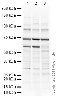
All lanes : Anti-SUZ12 antibody (ab12073) at 1 µg/ml
Lane 1 : SW480 (Human colon adenocarcinoma cell line) Whole Cell Lysate
Lane 2 : MCF7 (Human breast adenocarcinoma cell line) Whole Cell Lysate
Lane 3 : Caco 2 (Human colonic carcinoma cell line) Whole Cell Lysate
Lysates/proteins at 20 µg per lane.
Secondary
All lanes : Goat Anti-Rabbit IgG H&L (HRP) preadsorbed (ab97080) at 1/5000 dilution
Developed using the ECL technique.
Performed under reducing conditions.
Predicted band size: 83 kDa
Observed band size: 95 kDa why is the actual band size different from the predicted?
Additional bands at: 30 kDa, 53 kDa, 60 kDa, 75 kDa. We are unsure as to the identity of these extra bands.
Exposure time: 4 minutes
References (123)
ab12073 has been referenced in 123 publications.
- Jain P et al. PHF19 mediated regulation of proliferation and invasiveness in prostate cancer cells. Elife 9:N/A (2020). PubMed: 32155117
- Papoutsoglou P et al. The TGFB2-AS1 lncRNA Regulates TGF-ß Signaling by Modulating Corepressor Activity. Cell Rep 28:3182-3198.e11 (2019). PubMed: 31533040
- Xie H et al. LncRNA TRG-AS1 promotes glioblastoma cell proliferation by competitively binding with miR-877-5p to regulate SUZ12 expression. Pathol Res Pract 215:152476 (2019). PubMed: 31196742
- Lei I et al. SWI/SNF Component BAF250a Coordinates OCT4 and WNT Signaling Pathway to Control Cardiac Lineage Differentiation. Front Cell Dev Biol 7:358 (2019). PubMed: 32039194
- Paschos K et al. Requirement for PRC1 subunit BMI1 in host gene activation by Epstein-Barr virus protein EBNA3C. Nucleic Acids Res N/A:N/A (2019). PubMed: 30649516
Images
-
Lane 1: Wild-type HAP1 cell lysate (20 µg)
Lane 2: SUZ12 knockout HAP1 cell lysate (20 µg)
Lane 3: Caco2 cell lysate (20 µg)
Lane 4: MCF7 cell lysate (20 µg)
Lanes 1 - 4: Merged signal (red and green). Green - ab12073 observed at 95 kDa. Red - loading control, ab8245, observed at 37 kDa.ab12073 was shown to specifically recognize SUZ12 in wild-type HAP1 cells along with additional cross-reactive bands. No band was observed when SUZ12 knockout samples were used. Wild-type and SUZ12 knockout samples were subjected to SDS-PAGE. ab12073 and ab8245 (loading control to GAPDH) were diluted to 1µg/mL and 1/10,000 respectively and incubated overnight at 4°C. Blots were developed with Goat anti-Rabbit IgG H&L (IRDye® 800CW) preadsorbed (ab216773) and Goat anti-Mouse IgG H&L (IRDye® 680RD) preadsorbed (ab216776) secondary antibodies at 1/10 000 dilution for 1 h at room temperature before imaging.
-
All lanes : Anti-SUZ12 antibody (ab12073)
Lane 1 : Wild-type HeLa cell lysate
Lane 2 : SUZ12 knockout HeLa cell lysate
Lysates/proteins at 20 µg per lane.
Performed under reducing conditions.
Predicted band size: 83 kDa
Observed band size: 95 kDa why is the actual band size different from the predicted?Lanes 1- 2: Merged signal (red and green). Green - ab12073 observed at 95 kDa. Red - Anti-GAPDH antibody [6C5] - Loading Control (ab8245) observed at 37 kDa.
ab12073 was shown to react with SUZ12 in wild-type HeLa cells in western blot. The band observed in knockout cell line ab264983 (knockout cell lysate ab257721) lane below 95kDa may represent truncated forms and cleaved fragments. This has not been investigated further. Wild-type HeLa and SUZ12 knockout HeLa cell lysates were subjected to SDS-PAGE. Membrane was blocked for 1 hour at room temperature in 0.1% TBST with 3% non-fat dried milk. ab12073 and Anti-GAPDH antibody [6C5] - Loading Control (ab8245) were incubated overnight at 4°C at a 1 µg/ml and a 1 in 20000 dilution respectively. Blots were developed with Goat anti-Rabbit IgG H&L (IRDye®800CW) preadsorbed (ab216773) and Goat anti-Mouse IgG H&L (IRDye®680RD) preadsorbed (ab216776) secondary antibodies at 1 in 20000 dilution for 1 hour at room temperature before imaging.
-
All lanes : Anti-SUZ12 antibody (ab12073) at 1 µg/ml
Lane 1 : untransfected 293T cell lysate
Lane 2 : 293T cells transfected with 5ug HA-Suz12
Lysates/proteins at 20 µg per lane.
Secondary
All lanes : Goat polyclonal to Rabbit IgG H&L (HRP) (Dako) at 1/2000 dilution
Performed under reducing conditions.
Predicted band size: 83 kDa
Observed band size: 95 kDa why is the actual band size different from the predicted?
Additional bands at: 76 kDa. We are unsure as to the identity of these extra bands.
-
All lanes : Anti-SUZ12 antibody (ab12073) at 1 µg/ml
Lane 1 : SW480 (Human colon adenocarcinoma cell line) Whole Cell Lysate
Lane 2 : MCF7 (Human breast adenocarcinoma cell line) Whole Cell Lysate
Lane 3 : Caco 2 (Human colonic carcinoma cell line) Whole Cell Lysate
Lysates/proteins at 20 µg per lane.
Secondary
All lanes : Goat Anti-Rabbit IgG H&L (HRP) preadsorbed (ab97080) at 1/5000 dilution
Developed using the ECL technique.
Performed under reducing conditions.
Predicted band size: 83 kDa
Observed band size: 95 kDa why is the actual band size different from the predicted?
Additional bands at: 30 kDa, 53 kDa, 60 kDa, 75 kDa. We are unsure as to the identity of these extra bands.
Exposure time: 4 minutes


