Anti-SDHB antibody (ab151684)
")
Key features and details
- Rabbit polyclonal to SDHB
- Suitable for: WB, IHC-P
- Knockout validated
- Reacts with: Human
- Isotype: IgG
Overview
-
Product name
Anti-SDHB antibody
See all SDHB primary antibodies -
Description
Rabbit polyclonal to SDHB -
Host species
Rabbit -
Tested Applications & Species
See all applications and species dataApplication Species IHC-P HumanWB Human
-
Immunogen
Synthetic peptide corresponding to Human SDHB aa 1-100 conjugated to keyhole limpet haemocyanin.
Database link: P21912 -
Positive control
- This antibody gave a positive signal in both Human Heart tissue and Human Heart Mitochondrial lysates. This antibody gave a positive result in IHC in the following FFPE tissue: Normal human heart muscle.
Properties
-
Form
Liquid -
Storage instructions
Shipped at 4°C. Store at +4°C short term (1-2 weeks). Upon delivery aliquot. Store at -20°C or -80°C. Avoid freeze / thaw cycle. -
Storage buffer
pH: 7.40
Preservative: 0.02% Sodium azide
Constituent: PBS
Batches of this product that have a concentration Concentration information loading...
Purity
Immunogen affinity purifiedClonality
PolyclonalIsotype
IgGResearch areas
Associated products
-
Compatible Secondaries
-
Isotype control
Applications
The Abpromise guarantee
Our Abpromise guarantee covers the use of ab151684 in the following tested applications.
The application notes include recommended starting dilutions; optimal dilutions/concentrations should be determined by the end user.
GuaranteedTested applications are guaranteed to work and covered by our Abpromise guarantee.
PredictedPredicted to work for this combination of applications and species but not guaranteed.
IncompatibleDoes not work for this combination of applications and species.
Application Species IHC-P HumanWB HumanAll applications RabbitHorseCowChimpanzeeMacaque monkeyGorillaOrangutanApplication Abreviews Notes WB Use a concentration of 1 µg/ml. Detects a band of approximately 29 kDa (predicted molecular weight: 32 kDa).IHC-P Use a concentration of 5 µg/ml. Perform heat mediated antigen retrieval with citrate buffer pH 6 before commencing with IHC staining protocol.Notes WB
Use a concentration of 1 µg/ml. Detects a band of approximately 29 kDa (predicted molecular weight: 32 kDa).IHC-P
Use a concentration of 5 µg/ml. Perform heat mediated antigen retrieval with citrate buffer pH 6 before commencing with IHC staining protocol.Target
-
Function
Iron-sulfur protein (IP) subunit of succinate dehydrogenase (SDH) that is involved in complex II of the mitochondrial electron transport chain and is responsible for transferring electrons from succinate to ubiquinone (coenzyme Q). -
Pathway
Carbohydrate metabolism; tricarboxylic acid cycle; fumarate from succinate (eukaryal route): step 1/1. -
Involvement in disease
Defects in SDHB are a cause of susceptibility to pheochromocytoma (PCC) [MIM:171300]. A catecholamine-producing tumor of chromaffin tissue of the adrenal medulla or sympathetic paraganglia. The cardinal symptom, reflecting the increased secretion of epinephrine and norepinephrine, is hypertension, which may be persistent or intermittent.
Defects in SDHB are the cause of hereditary paragangliomas type 4 (PGL4) [MIM:115310]; also known as familial non-chromaffin paragangliomas type 4. Paragangliomas refer to rare and mostly benign tumors that arise from any component of the neuroendocrine system. PGL4 is characterized by the development of mostly benign, highly vascular, slow growing tumors in the head and neck. In the head and neck region, the carotid body is the largest of all paraganglia and is also the most common site of the tumors.
Defects in SDHB are a cause of paraganglioma and gastric stromal sarcoma (PGGSS) [MIM:606864]; also called Carney-Stratakis syndrome. Gastrointestinal stromal tumors may be sporadic or inherited in an autosomal dominant manner, alone or as a component of a syndrome associated with other tumors, such as in the context of neurofibromatosis type 1 (NF1). Patients have both gastrointestinal stromal tumors and paragangliomas. Susceptibility to the tumors was inherited in an apparently autosomal dominant manner, with incomplete penetrance.
Defects in SDHB are a cause of Cowden-like syndrome (CWDLS) [MIM:612359]. Cowden-like syndrome is a cancer predisposition syndrome associated with elevated risk for tumors of the breast, thyroid, kidney and uterus. -
Sequence similarities
Belongs to the succinate dehydrogenase/fumarate reductase iron-sulfur protein family.
Contains 1 2Fe-2S ferredoxin-type domain.
Contains 1 4Fe-4S ferredoxin-type domain. -
Cellular localization
Mitochondrion inner membrane. - Information by UniProt
-
Database links
- Entrez Gene: 286840 Cow
- Entrez Gene: 6390 Human
- Omim: 185470 Human
- SwissProt: Q3T189 Cow
- SwissProt: P21912 Human
- Unigene: 465924 Human
-
Alternative names
- CWS2 antibody
- DHSB_HUMAN antibody
- FLJ92337 antibody
see all
Images
-
Western blot - Anti-SDHB antibody (ab151684)All lanes : Anti-SDHB antibody (ab151684) at 1 µg/ml
Lane 1 : Wild-type HEK-293 whole cell lysate
Lane 2 : SDHB knockout HEK-293 whole cell lysate
Lane 3 : HepG2 whole cell lysate
Lysates/proteins at 20 µg per lane.
Predicted band size: 32 kDa
Observed band size: 32 kDaLanes 1 - 3: Merged signal (red and green). Green - ab151684 observed at 32 kDa. Red - loading control, ab130007, observed at 130 kDa.
ab151684 was shown to recognize SDHB in wild-type HEK-293 cells as signal was lost at the expected MW in SDHB knockout cells. Additional cross-reactive bands were observed in the wild-type and knockout cells. Wild-type and SDHB knockout samples were subjected to SDS-PAGE. Ab151684 and ab130007 (Mouse anti-Vinculin loading control) were incubated overnight at 4°C at 1 ug/ml and 1/20000 dilution respectively. Blots were developed with Goat anti-Rabbit IgG H&L (IRDye® 800CW) preabsorbed ab216773 and Goat anti-Mouse IgG H&L (IRDye® 680RD) preabsorbed ab216776 secondary antibodies at 1/20000 dilution for 1 hour at room temperature before imaging.
-
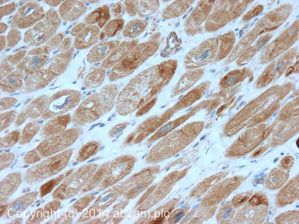 Immunohistochemistry (Formalin/PFA-fixed paraffin-embedded sections) - Anti-SDHB antibody (ab151684)
IHC image of SDHB staining in normal human heart muscle formalin fixed paraffin embedded tissue section, performed on a Leica Bond™ system using the standard protocol F. The section was pre-treated using heat mediated antigen retrieval with sodium citrate buffer (pH6, epitope retrieval solution 1) for 20 mins. The section was then incubated with ab151684, 5µg/ml, for 15 mins at room temperature and detected using an HRP conjugated compact polymer system. DAB was used as the chromogen. The section was then counterstained with haematoxylin and mounted with DPX.
For other IHC staining systems (automated and non-automated) customers should optimize variable parameters such as antigen retrieval conditions, primary antibody concentration and antibody incubation times.
-
 Western blot - Anti-SDHB antibody (ab151684)All lanes : Anti-SDHB antibody (ab151684) at 1 µg/ml
Lane 1 : Human heart tissue lysate - total protein (ab29431)
Lane 2 : Human Heart Mitochondrial Lysate
Lysates/proteins at 10 µg per lane.
Secondary
All lanes : Goat Anti-Rabbit IgG H&L (HRP) (ab97051) at 1/10000 dilution
Developed using the ECL technique.
Performed under reducing conditions.
Predicted band size: 32 kDa
Observed band size: 29 kDa why is the actual band size different from the predicted?
Exposure time: 1 minuteThe band observed at 29 kDa could potentially be a cleaved form of SDHB due to the presence of a 28 amino acid transit peptide.
This blot was produced using a 4-12% Bis-tris gel under the MOPS buffer system. The gel was run at 200V for 50 minutes before being transferred onto a Nitrocellulose membrane at 30V for 70 minutes. The membrane was then blocked for an hour using 5% Bovine Serum Albumin before being incubated with ab151684 overnight at 4°C. Antibody binding was detected using an anti-rabbit antibody conjugated to HRP, and visualised using ECL development solution.
Protocols
Datasheets and documents
-
SDS download
-
Datasheet download
References (1)
ab151684 has been referenced in 1 publication.
- Zhang Q et al. Deletion of Mtu1 (Trmu) in zebrafish revealed the essential role of tRNA modification in mitochondrial biogenesis and hearing function. Nucleic Acids Res 46:10930-10945 (2018). PubMed: 30137487
Images
-
Western blot - Anti-SDHB antibody (ab151684)All lanes : Anti-SDHB antibody (ab151684) at 1 µg/ml
Lane 1 : Wild-type HEK-293 whole cell lysate
Lane 2 : SDHB knockout HEK-293 whole cell lysate
Lane 3 : HepG2 whole cell lysate
Lysates/proteins at 20 µg per lane.
Predicted band size: 32 kDa
Observed band size: 32 kDaLanes 1 - 3: Merged signal (red and green). Green - ab151684 observed at 32 kDa. Red - loading control, ab130007, observed at 130 kDa.
ab151684 was shown to recognize SDHB in wild-type HEK-293 cells as signal was lost at the expected MW in SDHB knockout cells. Additional cross-reactive bands were observed in the wild-type and knockout cells. Wild-type and SDHB knockout samples were subjected to SDS-PAGE. Ab151684 and ab130007 (Mouse anti-Vinculin loading control) were incubated overnight at 4°C at 1 ug/ml and 1/20000 dilution respectively. Blots were developed with Goat anti-Rabbit IgG H&L (IRDye® 800CW) preabsorbed ab216773 and Goat anti-Mouse IgG H&L (IRDye® 680RD) preabsorbed ab216776 secondary antibodies at 1/20000 dilution for 1 hour at room temperature before imaging.
-
Immunohistochemistry (Formalin/PFA-fixed paraffin-embedded sections) - Anti-SDHB antibody (ab151684)
IHC image of SDHB staining in normal human heart muscle formalin fixed paraffin embedded tissue section, performed on a Leica Bond™ system using the standard protocol F. The section was pre-treated using heat mediated antigen retrieval with sodium citrate buffer (pH6, epitope retrieval solution 1) for 20 mins. The section was then incubated with ab151684, 5µg/ml, for 15 mins at room temperature and detected using an HRP conjugated compact polymer system. DAB was used as the chromogen. The section was then counterstained with haematoxylin and mounted with DPX.
For other IHC staining systems (automated and non-automated) customers should optimize variable parameters such as antigen retrieval conditions, primary antibody concentration and antibody incubation times.
-
Western blot - Anti-SDHB antibody (ab151684)All lanes : Anti-SDHB antibody (ab151684) at 1 µg/ml
Lane 1 : Human heart tissue lysate - total protein (ab29431)
Lane 2 : Human Heart Mitochondrial Lysate
Lysates/proteins at 10 µg per lane.
Secondary
All lanes : Goat Anti-Rabbit IgG H&L (HRP) (ab97051) at 1/10000 dilution
Developed using the ECL technique.
Performed under reducing conditions.
Predicted band size: 32 kDa
Observed band size: 29 kDa why is the actual band size different from the predicted?
Exposure time: 1 minuteThe band observed at 29 kDa could potentially be a cleaved form of SDHB due to the presence of a 28 amino acid transit peptide.
This blot was produced using a 4-12% Bis-tris gel under the MOPS buffer system. The gel was run at 200V for 50 minutes before being transferred onto a Nitrocellulose membrane at 30V for 70 minutes. The membrane was then blocked for an hour using 5% Bovine Serum Albumin before being incubated with ab151684 overnight at 4°C. Antibody binding was detected using an anti-rabbit antibody conjugated to HRP, and visualised using ECL development solution.


