Anti-SATB1 antibody (ab101085)
Key features and details
- Rabbit polyclonal to SATB1
- Suitable for: WB
- Knockout validated
- Reacts with: Human
- Isotype: IgG
Overview
-
Product name
Anti-SATB1 antibody
See all SATB1 primary antibodies -
Description
Rabbit polyclonal to SATB1 -
Host species
Rabbit -
Tested Applications & Species
See all applications and species dataApplication Species WB Human -
Immunogen
Synthetic peptide corresponding to Human SATB1 aa 550-650 conjugated to keyhole limpet haemocyanin.
(Peptide available asab111651) -
Positive control
- This antibody gave a positive signal in Jurkat whole cell lysate.
Properties
-
Form
Liquid -
Storage instructions
Shipped at 4°C. Store at +4°C short term (1-2 weeks). Upon delivery aliquot. Store at -20°C or -80°C. Avoid freeze / thaw cycle. -
Storage buffer
pH: 7.40
Preservative: 0.02% Sodium azide
Constituents: 1% BSA, PBS
Batches of this product that have a concentration Concentration information loading...
Purity
Immunogen affinity purifiedClonality
PolyclonalIsotype
IgGResearch areas
Associated products
-
Compatible Secondaries
-
Isotype control
Applications
The Abpromise guarantee
Our Abpromise guarantee covers the use of ab101085 in the following tested applications.
The application notes include recommended starting dilutions; optimal dilutions/concentrations should be determined by the end user.
GuaranteedTested applications are guaranteed to work and covered by our Abpromise guarantee.
PredictedPredicted to work for this combination of applications and species but not guaranteed.
IncompatibleDoes not work for this combination of applications and species.
Application Species WB HumanAll applications MouseRatCowDogPigApplication Abreviews Notes WB Use a concentration of 1 µg/ml. Detects a band of approximately 100 kDa (predicted molecular weight: 86 kDa).Notes WB
Use a concentration of 1 µg/ml. Detects a band of approximately 100 kDa (predicted molecular weight: 86 kDa).Target
-
Function
Crucial silencing factor contributing to the initiation of X inactivation mediated by Xist RNA that occurs during embryogenesis and in lymphoma (By similarity). Binds to DNA at special AT-rich sequences, the consensus SATB1-binding sequence (CSBS), at nuclear matrix- or scaffold-associated regions. Thought to recognize the sugar-phosphate structure of double-stranded DNA. Transcriptional repressor controlling nuclear and viral gene expression in a phosphorylated and acetylated status-dependent manner, by binding to matrix attachment regions (MARs) of DNA and inducing a local chromatin-loop remodeling. Acts as a docking site for several chromatin remodeling enzymes (e.g. PML at the MHC-I locus) and also by recruiting corepressors (HDACs) or coactivators (HATs) directly to promoters and enhancers. Modulates genes that are essential in the maturation of the immune T-cell CD8SP from thymocytes. Required for the switching of fetal globin species, and beta- and gamma-globin genes regulation during erythroid differentiation. Plays a role in chromatin organization and nuclear architecture during apoptosis. Interacts with the unique region (UR) of cytomegalovirus (CMV). Alu-like motifs and SATB1-binding sites provide a unique chromatin context which seems preferentially targeted by the HIV-1 integration machinery. Moreover, HIV-1 Tat may overcome SATB1-mediated repression of IL2 and IL2RA (interleukin) in T-cells by binding to the same domain than HDAC1. Delineates specific epigenetic modifications at target gene loci, directly upregulating metastasis-associated genes while downregulating tumor-suppressor genes. Reprograms chromatin organization and the transcription profiles of breast tumors to promote growth and metastasis. -
Tissue specificity
Expressed predominantly in thymus. -
Sequence similarities
Belongs to the CUT homeobox family.
Contains 2 CUT DNA-binding domains.
Contains 1 homeobox DNA-binding domain. -
Post-translational
modificationsSumoylated. Sumoylation promotes cleavage by caspases.
Phosphorylated by PKC. Acetylated by PCAF. Phosphorylated form interacts with HDAC1, but unphosphorylated form interacts with PCAF. DNA binding properties are activated by phosphorylation and inactivated by acetylation. In opposition, gene expression is down-regulated by phosphorylation but up-regulated by acetylation.
Cleaved at Asp-254 by caspase-3 and caspase-6 during T-cell apoptosis in thymus and during B-cell stimulation. The cleaved forms can not dimerize and lose transcription regulation function because of impaired DNA and chromatin association. -
Cellular localization
Nucleus matrix. Nucleus > PML body. Organized into a cage-like network anchoring loops of heterochromatin and tethering specialized DNA sequences. When sumoylated, localized in promyelocytic leukemia nuclear bodies. - Information by UniProt
-
Database links
- Entrez Gene: 6304 Human
- Entrez Gene: 20230 Mouse
- Entrez Gene: 316164 Rat
- Omim: 602075 Human
- SwissProt: Q01826 Human
- SwissProt: Q60611 Mouse
- Unigene: 517717 Human
- Unigene: 311655 Mouse
see all -
Form
There are 2 isoforms produced by alternative splicing. -
Alternative names
- DNA binding protein SATB1 antibody
- DNA-binding protein SATB1 antibody
- SATB homeobox 1 antibody
see all
Images
-
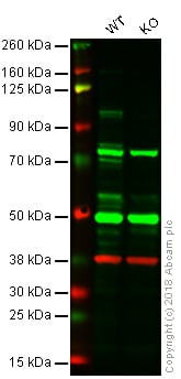
Lane 1: Wild-type HAP1 whole cell lysate (20 µg)
Lane 2: SATB1 knockout HAP1 whole cell lysate (20 µg)
Lanes 1 - 2: Merged signal (red and green). Green - ab101085 observed at 100 kDa. Red - loading control, ab9484, observed at 37 kDa.ab101085 was shown to recognize SATB1 in wild-type HAP1 cells as signal was lost at the expected MW in SATB1 knockout cells. Additional cross-reactive bands were observed in the wild-type and knockout cells. Wild-type and SATB1 knockout samples were subjected to SDS-PAGE. ab101085 and ab9484 (Mouse anti-GAPDH loading control) were incubated overnight at 4°C at 1 μg/ml and 1/20000 dilution respectively. Blots were developed with Goat anti-Rabbit IgG H&L (IRDye® 800CW) preabsorbed ab216773 and Goat anti-Mouse IgG H&L (IRDye® 680RD) preabsorbed ab216776 secondary antibodies at 1/20000 dilution for 1 hour at room temperature before imaging.
-
Anti-SATB1 antibody (ab101085) at 1 µg/ml + Jurkat (Human T cell lymphoblast-like cell line) Whole Cell Lysate at 10 µg
Secondary
Goat Anti-Rabbit IgG H&L (HRP) preadsorbed (ab97080) at 1/5000 dilution
Developed using the ECL technique.
Performed under reducing conditions.
Predicted band size: 86 kDa
Observed band size: 100 kDa why is the actual band size different from the predicted?
Additional bands at: 130 kDa, 45 kDa. We are unsure as to the identity of these extra bands.
Exposure time: 3 minutes
The predicted molecular weight of SATB1 is 86 kDa (SwissProt), however we expect to observe a banding pattern at 100 kDa. Abcam welcomes customer feedback and would appreciate any comments regarding this product and the data presented above.
Datasheets and documents
-
SDS download
-
Datasheet download
References (0)
ab101085 has not yet been referenced specifically in any publications.
Images
-
Lane 1: Wild-type HAP1 whole cell lysate (20 µg)
Lane 2: SATB1 knockout HAP1 whole cell lysate (20 µg)
Lanes 1 - 2: Merged signal (red and green). Green - ab101085 observed at 100 kDa. Red - loading control, ab9484, observed at 37 kDa.ab101085 was shown to recognize SATB1 in wild-type HAP1 cells as signal was lost at the expected MW in SATB1 knockout cells. Additional cross-reactive bands were observed in the wild-type and knockout cells. Wild-type and SATB1 knockout samples were subjected to SDS-PAGE. ab101085 and ab9484 (Mouse anti-GAPDH loading control) were incubated overnight at 4°C at 1 μg/ml and 1/20000 dilution respectively. Blots were developed with Goat anti-Rabbit IgG H&L (IRDye® 800CW) preabsorbed ab216773 and Goat anti-Mouse IgG H&L (IRDye® 680RD) preabsorbed ab216776 secondary antibodies at 1/20000 dilution for 1 hour at room temperature before imaging.
-
Anti-SATB1 antibody (ab101085) at 1 µg/ml + Jurkat (Human T cell lymphoblast-like cell line) Whole Cell Lysate at 10 µg
Secondary
Goat Anti-Rabbit IgG H&L (HRP) preadsorbed (ab97080) at 1/5000 dilution
Developed using the ECL technique.
Performed under reducing conditions.
Predicted band size: 86 kDa
Observed band size: 100 kDa why is the actual band size different from the predicted?
Additional bands at: 130 kDa, 45 kDa. We are unsure as to the identity of these extra bands.
Exposure time: 3 minutes
The predicted molecular weight of SATB1 is 86 kDa (SwissProt), however we expect to observe a banding pattern at 100 kDa. Abcam welcomes customer feedback and would appreciate any comments regarding this product and the data presented above.


