Anti-RhoA antibody (ab86297)
Key features and details
- Rabbit polyclonal to RhoA
- Suitable for: ICC/IF, WB
- Reacts with: Mouse, Rat, Human
- Isotype: IgG
Overview
-
Product name
Anti-RhoA antibody
See all RhoA primary antibodies -
Description
Rabbit polyclonal to RhoA -
Host species
Rabbit -
Tested applications
Suitable for: ICC/IF, WBmore details -
Species reactivity
Reacts with: Mouse, Rat, Human
Predicted to work with: Chicken, Cow, Dog, Zebrafish, Orangutan
-
Immunogen
Synthetic peptide corresponding to Human RhoA aa 150 to the C-terminus conjugated to keyhole limpet haemocyanin.
(Peptide available asab101307) -
Positive control
- This antibody gave a positive signal in the following whole cell lysates: HL60; MCF7; MDA-MB-231; HeLa; Human Platelets.
-
General notes
Reproducibility is key to advancing scientific discovery and accelerating scientists’ next breakthrough.
Abcam is leading the way with our range of recombinant antibodies, knockout-validated antibodies and knockout cell lines, all of which support improved reproducibility.
We are also planning to innovate the way in which we present recommended applications and species on our product datasheets, so that only applications & species that have been tested in our own labs, our suppliers or by selected trusted collaborators are covered by our Abpromise™ guarantee.
In preparation for this, we have started to update the applications & species that this product is Abpromise guaranteed for.
We are also updating the applications & species that this product has been “predicted to work with,” however this information is not covered by our Abpromise guarantee.
Applications & species from publications and Abreviews that have not been tested in our own labs or in those of our suppliers are not covered by the Abpromise guarantee.
Please check that this product meets your needs before purchasing. If you have any questions, special requirements or concerns, please send us an inquiry and/or contact our Support team ahead of purchase. Recommended alternatives for this product can be found below, as well as customer reviews and Q&As.
Properties
-
Form
Liquid -
Storage instructions
Shipped at 4°C. Store at +4°C short term (1-2 weeks). Upon delivery aliquot. Store at -20°C or -80°C. Avoid freeze / thaw cycle. -
Storage buffer
pH: 7.40
Preservative: 0.02% Sodium azide
Constituent: PBS
Batches of this product that have a concentration Concentration information loading...
Purity
Immunogen affinity purifiedClonality
PolyclonalIsotype
IgGResearch areas
Associated products
-
Compatible Secondaries
-
Isotype control
-
Recombinant Protein
Applications
Our Abpromise guarantee covers the use of ab86297 in the following tested applications.
The application notes include recommended starting dilutions; optimal dilutions/concentrations should be determined by the end user.
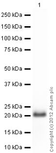
Application Abreviews Notes ICC/IF Use a concentration of 10 µg/ml. WB Use a concentration of 1 µg/ml. Detects a band of approximately 20 kDa (predicted molecular weight: 21 kDa). Target
-
Function
Regulates a signal transduction pathway linking plasma membrane receptors to the assembly of focal adhesions and actin stress fibers. Serves as a target for the yopT cysteine peptidase from Yersinia pestis, vector of the plague, and Yersinia pseudotuberculosis, which causes gastrointestinal disorders. May be an activator of PLCE1. Activated by ARHGEF2, which promotes the exchange of GDP for GTP. -
Sequence similarities
Belongs to the small GTPase superfamily. Rho family. -
Domain
The basic-rich region is essential for yopT recognition and cleavage. -
Post-translational
modificationsSubstrate for botulinum ADP-ribosyltransferase.
Cleaved by yopT protease when the cell is infected by some Yersinia pathogens. This removes the lipid attachment, and leads to its displacement from plasma membrane and to subsequent cytoskeleton cleavage.
AMPylation at Tyr-34 and Thr-37 are mediated by bacterial enzymes in case of infection by H.somnus and V.parahaemolyticus, respectively. AMPylation occurs in the effector region and leads to inactivation of the GTPase activity by preventing the interaction with downstream effectors, thereby inhibiting actin assembly in infected cells. It is unclear whether some human enzyme mediates AMPylation; FICD has such ability in vitro but additional experiments remain to be done to confirm results in vivo.
Ubiquitinated by the BCR(BACURD1) and BCR(BACURD2) E3 ubiquitin ligase complexes, leading to its degradation by the proteasome, thereby regulating the actin cytoskeleton and cell migration. -
Cellular localization
Cell membrane. Cytoplasm > cytoskeleton. - Information by UniProt
-
Database links
- Entrez Gene: 395442 Chicken
- Entrez Gene: 338049 Cow
- Entrez Gene: 403954 Dog
- Entrez Gene: 387 Human
- Entrez Gene: 11848 Mouse
- Entrez Gene: 117273 Rat
- Omim: 165390 Human
- SwissProt: P61585 Cow
see all -
Alternative names
- Aplysia ras related homolog 12 antibody
- ARH12 antibody
- ARHA antibody
see all
Images
-
All lanes : Anti-RhoA antibody (ab86297) at 1 µg/ml
Lane 1 : HL60 (Human promyelocytic leukemia cell line) Whole Cell Lysate
Lane 2 : MCF7 (Human breast adenocarcinoma cell line) Whole Cell Lysate
Lane 3 : MDA-MB-231 (Human breast adenocarcinoma cell line) Whole Cell Lysate
Lane 4 : HeLa (Human epithelial carcinoma cell line) Whole Cell Lysate
Lane 5 : Human Platelet (Human adult normal cell line) Whole Cell Lysate
Lysates/proteins at 10 µg per lane.
Secondary
All lanes : Goat Anti-Rabbit IgG H&L (HRP) preadsorbed (ab97080) at 1/5000 dilution
Developed using the ECL technique.
Performed under reducing conditions.
Predicted band size: 21 kDa
Observed band size: 20 kDa why is the actual band size different from the predicted?
Exposure time: 90 seconds -
All lanes : Anti-RhoA antibody (ab86297) at 1 µg/ml
Lane 1 : Kidney (Mouse) Tissue Lysate
Lane 2 :Mouse lung normal tissue lysate - total protein (ab29297)
Lane 3 : Kidney (Rat) Tissue Lysate
Lane 4 : Lung (Rat) Tissue Lysate
Lysates/proteins at 10 µg per lane.
Secondary
All lanes : Goat Anti-Rabbit IgG H&L (HRP) preadsorbed (ab97080) at 1/5000 dilution
Developed using the ECL technique.
Performed under reducing conditions.
Predicted band size: 21 kDa
Observed band size: 20 kDa why is the actual band size different from the predicted?
Exposure time: 2 minutes -
ICC/IF image of ab86297 stained MCF7 cells. The cells were 4% PFA fixed (10 min) and then incubated in 1%BSA / 10% normal goat serum / 0.3M glycine in 0.1% PBS-Tween for 1h to permeabilise the cells and block non-specific protein-protein interactions. The cells were then incubated with the antibody (ab86297, 10µg/ml) overnight at +4°C. The secondary antibody (green) was ab96899 Dylight 488 goat anti-rabbit IgG (H+L) used at a 1/250 dilution for 1h. Alexa Fluor® 594 WGA was used to label plasma membranes (red) at a 1/200 dilution for 1h. DAPI was used to stain the cell nuclei (blue) at a concentration of 1.43µM.
Protocols
Datasheets and documents
References (8)
ab86297 has been referenced in 8 publications.
- Fan XD et al. miR-154-3p and miR-487-3p synergistically modulate RHOA signaling in the carcinogenesis of thyroid cancer. Biosci Rep 40:N/A (2020). PubMed: 31820783
- Abou-Antoun TJ et al. Molecular and functional analysis of anchorage independent, treatment-evasive neuroblastoma tumorspheres with enhanced malignant properties: A possible explanation for radio-therapy resistance. PLoS One 13:e0189711 (2018). PubMed: 29298329
- Ye Y et al. Exosomal miR-141-3p regulates osteoblast activity to promote the osteoblastic metastasis of prostate cancer. Oncotarget 8:94834-94849 (2017). PubMed: 29212270
- Wang Y et al. MiR-124 Promote Neurogenic Transdifferentiation of Adipose Derived Mesenchymal Stromal Cells Partly through RhoA/ROCK1, but Not ROCK2 Signaling Pathway. PLoS One 11:e0146646 (2016). WB ; Human . PubMed: 26745800
- Lin R et al. Electroacupuncture improves cognitive function through Rho GTPases and enhances dendritic spine plasticity in rats with cerebral ischemia-reperfusion. Mol Med Rep 13:2655-60 (2016). PubMed: 26846874
- Freeman MC et al. Coronaviruses induce entry-independent, continuous macropinocytosis. MBio 5:e01340-14 (2014). Mouse . PubMed: 25096879
- Lázaro-Diéguez F et al. Par1b links lumen polarity with LGN-NuMA positioning for distinct epithelial cell division phenotypes. J Cell Biol 203:251-64 (2013). PubMed: 24165937
- Nuno DW et al. RhoA localization with caveolin-1 regulates vascular contractions to serotonin. Am J Physiol Regul Integr Comp Physiol 303:R959-67 (2012). WB ; Mouse . PubMed: 22955057
Images
-
All lanes : Anti-RhoA antibody (ab86297) at 1 µg/ml
Lane 1 : HL60 (Human promyelocytic leukemia cell line) Whole Cell Lysate
Lane 2 : MCF7 (Human breast adenocarcinoma cell line) Whole Cell Lysate
Lane 3 : MDA-MB-231 (Human breast adenocarcinoma cell line) Whole Cell Lysate
Lane 4 : HeLa (Human epithelial carcinoma cell line) Whole Cell Lysate
Lane 5 : Human Platelet (Human adult normal cell line) Whole Cell Lysate
Lysates/proteins at 10 µg per lane.
Secondary
All lanes : Goat Anti-Rabbit IgG H&L (HRP) preadsorbed (ab97080) at 1/5000 dilution
Developed using the ECL technique.
Performed under reducing conditions.
Predicted band size: 21 kDa
Observed band size: 20 kDa why is the actual band size different from the predicted?
Exposure time: 90 seconds
-
All lanes : Anti-RhoA antibody (ab86297) at 1 µg/ml
Lane 1 : Kidney (Mouse) Tissue Lysate
Lane 2 :Mouse lung normal tissue lysate - total protein (ab29297)
Lane 3 : Kidney (Rat) Tissue Lysate
Lane 4 : Lung (Rat) Tissue Lysate
Lysates/proteins at 10 µg per lane.
Secondary
All lanes : Goat Anti-Rabbit IgG H&L (HRP) preadsorbed (ab97080) at 1/5000 dilution
Developed using the ECL technique.
Performed under reducing conditions.
Predicted band size: 21 kDa
Observed band size: 20 kDa why is the actual band size different from the predicted?
Exposure time: 2 minutes -
-
ICC/IF image of ab86297 stained MCF7 cells. The cells were 4% PFA fixed (10 min) and then incubated in 1%BSA / 10% normal goat serum / 0.3M glycine in 0.1% PBS-Tween for 1h to permeabilise the cells and block non-specific protein-protein interactions. The cells were then incubated with the antibody (ab86297, 10µg/ml) overnight at +4°C. The secondary antibody (green) was ab96899 Dylight 488 goat anti-rabbit IgG (H+L) used at a 1/250 dilution for 1h. Alexa Fluor® 594 WGA was used to label plasma membranes (red) at a 1/200 dilution for 1h. DAPI was used to stain the cell nuclei (blue) at a concentration of 1.43µM.


