Anti-PU.1/Spi1 antibody (ab88082)
")
Key features and details
- Mouse monoclonal to PU.1/Spi1
- Suitable for: WB, ELISA, ICC/IF, Flow Cyt
- Reacts with: Mouse, Human, Recombinant fragment
- Isotype: IgG1
Overview
-
Product name
Anti-PU.1/Spi1 antibody
See all PU.1/Spi1 primary antibodies -
Description
Mouse monoclonal to PU.1/Spi1 -
Host species
Mouse -
Tested Applications & Species
See all applications and species dataApplication Species ELISA Recombinant fragmentFlow Cyt HumanICC/IF HumanWB MouseHuman
-
Immunogen
Recombinant fragment, corresponding to amino acids 7-126 of human PU.1/Spi1 (NP_003111) with a proprietary tag.
-
Positive control
- Purchase matching WB positive control:Recombinant Human PU.1/Spi1 protein
- HeLa cells; Raw 264.7 cell lysate.
-
General notes
This product was changed from ascites to tissue culture supernatant on 15 May 2019. Please note that the dilutions may need to be adjusted accordingly. If you have any questions, please do not hesitate to contact our scientific support team.
The Life Science industry has been in the grips of a reproducibility crisis for a number of years. Abcam is leading the way in addressing the problem with our range of recombinant monoclonal antibodies and knockout edited cell lines for gold-standard validation.
One factor contributing to the crisis is the use of antibodies that are not suitable. This can lead to misleading results and the use of incorrect data informing project assumptions and direction. To help address this challenge, we have introduced an application and species grid on our primary antibody datasheets to make it easy to simplify identification of the right antibody for your needs.
Learn more here.
Properties
-
Form
Liquid -
Storage instructions
Shipped at 4°C. Upon delivery aliquot and store at -20°C or -80°C. Avoid repeated freeze / thaw cycles. -
Storage buffer
pH: 7.4
Constituent: 2.68% PBS -
 Concentration information loading...
 -
Purity
Tissue culture supernatant -
Purification notes
Purified from TCS. -
Clonality
Monoclonal -
Isotype
IgG1 -
Light chain type
kappa -
Research areas
Images
-
Western blot - Anti-PU.1/Spi1 antibody (ab88082)Anti-PU.1/Spi1 antibody (ab88082) at 5 µg/ml + tagged human recombinant protein fragment (the immunogen) at 0.2 µg
Secondary
Goat Anti-Mouse IgG (H&L)-HRP Conjugate at 1/5000 dilution
Predicted band size: 31 kDa
Observed band size: 39 kDa why is the actual band size different from the predicted?Note: Molecular weight of the proprietary tag alone is 26 kDa.
This image was generated using the ascites version of the product.
-
 Immunocytochemistry/ Immunofluorescence - Anti-PU.1/Spi1 antibody (ab88082)
ab88082, at 10 µg/ml, staining PU.1/Spi1 in HeLa cells by Immunofluorescence.
This image was generated using the ascites version of the product.
-
 ELISA - Anti-PU.1/Spi1 antibody (ab88082)
Detection limit for recombinant (human) tagged ab88082 is approximately 0.1ng/ml as a capture antibody.
This image was generated using the ascites version of the product.
-
 Western blot - Anti-PU.1/Spi1 antibody (ab88082)Anti-PU.1/Spi1 antibody (ab88082) at 5 µg/ml + Raw 264.7 cell lysate (Mouse) at 25 µg
Secondary
Goat anti-Mouse IgG (H&L)-HRP at 1/2500 dilution
Predicted band size: 31 kDa
Observed band size: 31 kDaThis image was generated using the ascites version of the product.
-
 Immunocytochemistry/ Immunofluorescence - Anti-PU.1/Spi1 antibody (ab88082) Image courtesy of Sellgren et al. Mol Psychiatry. 2017 Feb; 22(2): 170–177. Fig S3: Doi: 10.1038/mp.2016.220
Ab88082 staining PU.1/Spi1 in human induced microglia by ICC/IF (Immunocytochemistry/Immunofluorescence). Cells were fixed with 4% paraformaldehyde for 15 minutes. Samples were incubated with primary antibody (1:200) for 2 hours at room temperature. Hoechst nuclear stain (blue) was used at 1:5000 dilution.
This image was generated using the ascites version of the product.
-
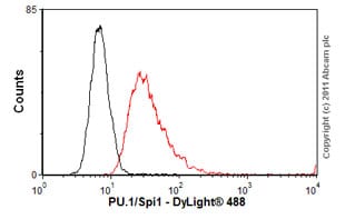 Flow Cytometry - Anti-PU.1/Spi1 antibody (ab88082)
Overlay histogram showing K562 cells stained with ab88082 (red line). The cells were fixed with 80% methanol (5 min) and then permeabilized with 0.1% PBS-Tween for 20 min. The cells were then incubated in 1x PBS / 10% normal goat serum / 0.3M glycine to block non-specific protein-protein interactions followed by the antibody (ab88082, 2µg/1x106 cells) for 30 min at 22ºC. The secondary antibody used was DyLight® 488 goat anti-mouse IgG (H+L) (ab96879) at 1/500 dilution for 30 min at 22ºC. Isotype control antibody (black line) was mouse IgG1 [ICIGG1] (ab91353, 2µg/1x106 cells) used under the same conditions. Acquisition of >5,000 events was performed.
This image was generated using the ascites version of the product.


