Anti-PCNA antibody (ab18197)
")
Key features and details
- Rabbit polyclonal to PCNA
- Suitable for: WB, ICC/IF, Flow Cyt, IHC-P
- Reacts with: Mouse, Rat, Human, Common marmoset
- Isotype: IgG
Overview
-
Product name
Anti-PCNA antibody
See all PCNA primary antibodies -
Description
Rabbit polyclonal to PCNA -
Host species
Rabbit -
Tested Applications & Species
See all applications and species dataApplication Species Flow Cyt HumanICC/IF MouseHumanIHC-P MouseCommon marmosetWB Human
-
Immunogen
Images
-
Western blot - Anti-PCNA antibody (ab18197)All lanes : Anti-PCNA antibody (ab18197) at 1 µg/ml
Lane 1 : HEK293 (Human embryonic kidney cell line) Whole Cell Lysate
Lane 2 : NIH 3T3 (Mouse embryonic fibroblast cell line) Whole Cell Lysate
Lane 3 : PC12 (Rat adrenal pheochromocytoma cell line) Whole Cell Lysate
Lane 4 : HEK293 (Human embryonic kidney cell line) Whole Cell Lysate with Human PCNA peptide (ab18602) at 1 µg/ml
Lane 5 : NIH 3T3 (Mouse embryonic fibroblast cell line) Whole Cell Lysate with Human PCNA peptide (ab18602) at 1 µg/ml
Lane 6 : PC12 (Rat adrenal pheochromocytoma cell line) Whole Cell Lysate with Human PCNA peptide (ab18602) at 1 µg/ml
Lysates/proteins at 20 µg per lane.
Secondary
All lanes : Goat Anti-Rabbit IgG H&L (HRP) preadsorbed (ab97080) at 1/5000 dilution
Developed using the ECL technique.
Performed under reducing conditions.
Predicted band size: 29 kDa
Observed band size: 29 kDa
Additional bands at: 48 kDa, 52 kDa. We are unsure as to the identity of these extra bands.
Exposure time: 1 minute
-
 Immunohistochemistry (Formalin/PFA-fixed paraffin-embedded sections) - Anti-PCNA antibody (ab18197) Image courtesy of Carl Hobbs, Kings College London, U.K.
ab18197 staining PCNA in tissue sections of the goat spleen by Immunohistochemistry (Formalin/PFA-fixed paraffin-embedded sections). Tissue was fixed with formaldehyde and a heat mediated antigen retrieval step was performed. Samples were then blocked with 1% B.S.A. for 10 minutes at 21ºC followed by incubation with the primary antibody for 2 hours at 1/4000. A biotin-conjugated goat anti-rabbit polyclonal was used as secondary antibody at a 1/250 dilution.
-
 Immunocytochemistry/ Immunofluorescence - Anti-PCNA antibody (ab18197) This image is courtesy of an Abreview submitted by Dr Kirk McManusab18197, at a 1/5000 dilution, staining PCNA in assynchronous HeLa cells. Cells were counter-stained with DAPI (red). For more information please refer to Abreview.
 -
 Flow Cytometry - Anti-PCNA antibody (ab18197)
Overlay histogram showing HeLa cells stained with ab18197 (red line). The cells were fixed with 80% methanol (5 min) and then permeabilized with 0.1% PBS-Tween 20 for 20 min. The cells were then incubated in 1x PBS / 10% normal goat serum / 0.3M glycine to block non-specific protein-protein interactions followed by the antibody (ab18197, 0.05μg/1x106 cells) for 30 min at 22ºC. The secondary antibody used was Alexa Fluor® 488 goat anti-rabbit IgG (H&L) (ab150081) at 1/4000 dilution for 30 min at 22ºC. Isotype control antibody (black line) was rabbit IgG (polyclonal) (ab171870, 0.05μg/1x106 cells) used under the same conditions. Unlabelled sample (blue line) was also used as a control.
Acquisition of >5,000 events were collected using a 20mW Argon ion laser (488nm) and 525/30 bandpass filter.
-
 Immunohistochemistry (Formalin/PFA-fixed paraffin-embedded sections) - Anti-PCNA antibody (ab18197) Image courtesy of Carl Hobbs, Kings College London, U.K.
ab18197 staining PCNA in tissue sections of the marmoset spleen by Immunohistochemistry (Formalin/PFA-fixed paraffin-embedded sections). Tissue was fixed with formaldehyde and a heat mediated antigen retrieval step was performed. Samples were then blocked with 1% B.S.A. for 10 minutes at 21ºC followed by incubation with the primary antibody for 2 hours at 1/6000. A biotin-conjugated goat anti-rabbit polyclonal was used as secondary antibody at a 1/250 dilution.
-
 Immunohistochemistry (Formalin/PFA-fixed paraffin-embedded sections) - Anti-PCNA antibody (ab18197) Image courtesy of Carl Hobbs, King College London, U.K.
ab18197 staining PCNA in tissue sections of the cow spleen by Immunohistochemistry (Formalin/PFA-fixed paraffin-embedded sections). Tissue was fixed with formaldehyde and a heat mediated antigen retrieval step was performed. Samples were then blocked with 1% B.S.A. for 10 minutes at 21ºC followed by incubation with the primary antibody for 2 hours at 1/4000. A biotin-conjugated goat anti-rabbit polyclonal was used as secondary antibody at a 1/250 dilution.
-
 Immunohistochemistry (Formalin/PFA-fixed paraffin-embedded sections) - Anti-PCNA antibody (ab18197) Image courtesy of Carl Hobbs, King College London, U.K.
ab18197 staining PCNA in tissue sections of the sheep spleen by Immunohistochemistry (Formalin/PFA-fixed paraffin-embedded sections). Tissue was fixed with formaldehyde and a heat mediated antigen retrieval step was performed. Samples were then blocked with 1% B.S.A. for 10 minutes at 21ºC followed by incubation with the primary antibody for 2 hours at 1/6000. A biotin-conjugated goat anti-rabbit polyclonal was used as secondary antibody at a 1/250 dilution.
-
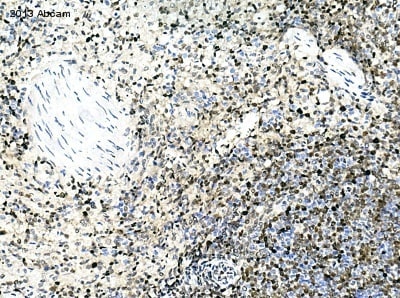
 Immunohistochemistry (Formalin/PFA-fixed paraffin-embedded sections) - Anti-PCNA antibody (ab18197) Image courtesy of Carl Hobbs, Kings College London, U.K.
ab18197 staining PCNA in tissue sections of the rat brain by Immunohistochemistry (Formalin/PFA-fixed paraffin-embedded sections). Tissue was fixed with formaldehyde and a heat mediated antigen retrieval step was performed. Samples were then blocked with 1% B.S.A. for 10 minutes at 21ºC followed by incubation with the primary antibody for 2 hours at 1/10000. A biotin-conjugated goat anti-rabbit polyclonal was used as secondary antibody at a 1/250 dilution.
-
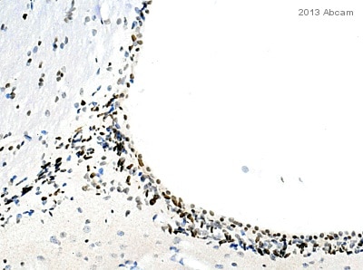
 Immunohistochemistry (Formalin/PFA-fixed paraffin-embedded sections) - Anti-PCNA antibody (ab18197) Image courtesy of Carl Hobbs, Kings College London, U.K.
ab18197 staining PCNA in tissue sections of the mouse brain by Immunohistochemistry (Formalin/PFA-fixed paraffin-embedded sections). Tissue was fixed with formaldehyde and a heat mediated antigen retrieval step was performed. Samples were then blocked with 1% B.S.A. for 10 minutes at 21ºC followed by incubation with the primary antibody for 2 hours at 1/6000. A biotin-conjugated goat anti-rabbit polyclonal was used as secondary antibody at a 1/250 dilution.
-
 Immunohistochemistry (Formalin/PFA-fixed paraffin-embedded sections) - Anti-PCNA antibody (ab18197) Image Courtesy of Carl Hobbs, Kings College London, U.K.
ab18197 staining PCNA in monkey COS cell pellet by Immunohistochemistry (Formalin/PFA-fixed paraffin-embedded sections). Tissue was fixed with formaldehyde and a heat mediated antigen retrieval step was performed. Samples were then blocked with 1% B.S.A. for 10 minutes at 21ºC followed by incubation with the primary antibody for 2 hours at 1/4000. A biotin-conjugated goat anti-rabbit polyclonal was used as secondary antibody at a 1/250 dilution.
-
 Immunocytochemistry/ Immunofluorescence - Anti-PCNA antibody (ab18197)
ICC/IF image of ab18197 stained HeLa cells. The cells were 100% methanol fixed (5 min) and then incubated in 1%BSA / 10% normal goat serum / 0.3M glycine in 0.1% PBS-Tween for 1h to permeabilise the cells and block non-specific protein-protein interactions. The cells were then incubated with the antibody (ab18197, 1µg/ml) overnight at +4°C. The secondary antibody (green) was DyLight® 488 goat anti-rabbit IgG - H&L, pre-adsorbed (ab96899) used at a 1/250 dilution for 1h. Alexa Fluor® 594 WGA was used to label plasma membranes (red) at a 1/200 dilution for 1h. DAPI was used to stain the cell nuclei (blue) at a concentration of 1.43µM.
-
 Immunocytochemistry/ Immunofluorescence - Anti-PCNA antibody (ab18197) Image courtesy of Hank Farr by Abreview.ab18197 staining PCNA in Zebrafish gastrula embryos by Immunocytochemistry/ Immunofluorescence (wholemount).
Zebrafish embryos were fixed overnight at 4°C when they had reached 60% epiboly. Cells were fixed in formaldehyde, permeabilized using Proteinase K, blocked with 2% goat serum for 2 hours at 20°C and then incubated with ab18197 at a 1/500 dilution for 16 hours at 4°C. The secondary used was an Alexa-Fluor 488 conjugated goat anti-rabbit polyclonal used at a 1/500 dilution. Cells were post-fixed in PFA for 20 minutes at room temperature after extensive washing of the secondary antibody.
The left panel shows DAPI stained nuclei, the center panel is PCNA staining, and the right panel is the merged image. -
 Immunocytochemistry/ Immunofluorescence - Anti-PCNA antibody (ab18197)
ICC/IF image of ab18197 stained NIH/3T3 cells. The cells were methanol fixed (5 min) and incubated with the antibody (ab18197, 1µg/ml) for 1h at room temperature. The secondary antibody (green) was Alexa Fluor® 488 goat anti-rabbit IgG (H+L) used at a 1/1000 dilution for 1h. Image-iTTM FX Signal Enhancer was used as the primary blocking agent, 5% BSA (in TBS-T) was used for all other blocking steps. DAPI was used to stain the cell nuclei (blue). Alexa Fluor® 594 WGA was used to label plasma membranes (red).
Panel A does not show the Alexa Fluor® 488 channel, Panel B shows the specfic nuclear staining by ab18197. -
 Immunocytochemistry/ Immunofluorescence - Anti-PCNA antibody (ab18197) This image is courtesy of an abreview submitted by Dr Catherine Greenab18197, at a 1/2000 dilution staining PCNA in MRC5 Sv40 transformed fibroblasts. Cells were counterstained with DAPI (blue). For more information please refer to Abreview.
 -
 Immunocytochemistry/ Immunofluorescence - Anti-PCNA antibody (ab18197)ab18197 staining PCNA in SK-N-SH cells treated with KN-62 (ab120421), by ICC/IF. Increase in PCNA nuclear expression correlates with increased concentration of KN-62, as described in literature.
The cells were incubated at 37øC for 24h in media containing different concentrations of ab120421 (KN-62) in DMSO, fixed with 100% methanol for 5 minutes at -20øC and blocked with PBS containing 10% goat serum, 0.3 M glycine, 1% BSA and 0.1% tween for 2h at room temperature. Staining of the treated cells with ab18197 (1 æg/ml) was performed overnight at 4øC in PBS containing 1% BSA and 0.1% tween. A DyLight 488 goat anti-rabbit polyclonal antibody (ab96899) at 1/250 dilution was used as the secondary antibody. Nuclei were counterstained with DAPI and are shown in blue.
-
 Western blot - Anti-PCNA antibody (ab18197)


