Anti-p53 antibody (ab232930)
")
Key features and details
- Rabbit polyclonal to p53
- Suitable for: WB, ChIP, ChIP-sequencing
- Reacts with: Human
- Isotype: IgG
Overview
-
Product name
Anti-p53 antibody
See all p53 primary antibodies -
Description
Rabbit polyclonal to p53 -
Host species
Rabbit -
Tested Applications & Species
See all applications and species dataApplication Species ChIP HumanChIP-seq HumanWB Human
-
Immunogen
Synthetic peptide corresponding to Human p53 (C terminal) conjugated to keyhole limpet haemocyanin.
Database link: P04637 -
Positive control
- ChIP: Chromatin from U-2 OS cells treated with camptothecin. ChIPseq: Chromatin from U-2 OS cells. WB: HeLa nuclear extract.
Properties
-
Form
Liquid -
Storage instructions
Shipped at 4°C. Store at +4°C short term (1-2 weeks). Upon delivery aliquot. Store at -20°C long term. Avoid freeze / thaw cycle. -
Storage buffer
Preservatives: 0.05% Sodium azide, 0.05% Proclin 300
Constituent: PBS -
 Concentration information loading...
 -
Purity
Affinity purified -
Clonality
Polyclonal -
Isotype
IgG -
Research areas
Images
-
Western blot - Anti-p53 antibody (ab232930)Anti-p53 antibody (ab232930) at 1/2000 dilution + HeLa (human epithelial cell line from cervix adenocarcinoma) nuclear extract at 40 µg
Predicted band size: 44 kDa
-
 ChIP - Anti-p53 antibody (ab232930)
ChIP assays were performed using U-2 OS cells, treated with camptothecin, the ab232930 against p53 and optimized PCR primer sets for QPCR. ChIP was performed on sheared chromatin from 4 million cells. A titration of ab232930 consisting of 1, 2, 5, and 10 μg per ChIP experiment was analysed. IgG (2 μg/IP) was used as negative IP control. QPCR was performed with primers for the p21 and GAS6 genes used as positive controls, and for GAPDH gene and the Sat2 satellite repeat, used as negative controls. The figure shows the recovery, expressed as a % of input (the relative amount of immunoprecipitated DNA compared to input DNA after qPCR analysis).
-
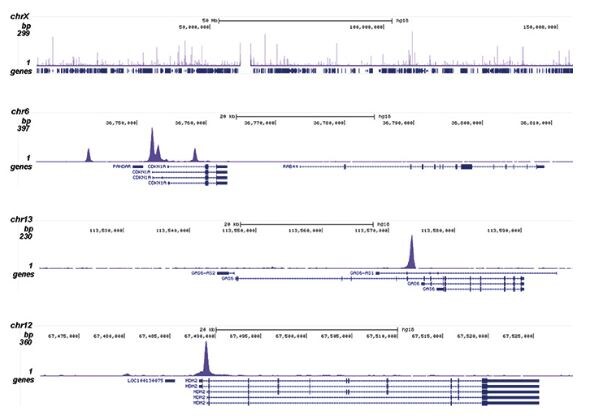 ChIP-sequencing - Anti-p53 antibody (ab232930)
ChIP was performed on sheared chromatin from 4 million U-2 OS (human bone osteosarcoma epithelial cell line) cells using 1 μg of ab232930 against p53. The IP’d DNA was subsequently analysed on an Illumina HiSeq. Library preparation, cluster generation and sequencing were performed according to the manufacturer’s instructions. The 51 bp tags were aligned to the human genome using the BWA algorithm. The figure shows the peak distribution along the X-chromosome (A) and in 3 genomic regions of chromosome 6, 13 and 12, surrounding p21 (CDKN1A), GAS6 and MDM2, 3 known targets genes of p53 (B, C and D, respectively).


