Anti-Spt6 antibody (ab32820)
Key features and details
- Rabbit polyclonal to Spt6
- Suitable for: IP, WB
- Reacts with: Human
- Isotype: IgG
Overview
-
Product name
Anti-Spt6 antibody
See all Spt6 primary antibodies -
Description
Rabbit polyclonal to Spt6 -
Host species
Rabbit -
Tested Applications & Species
See all applications and species dataApplication Species IP HumanWB Human -
Immunogen
-
Positive control
- This antibody gave a positive signal for the following cell lysates: HeLa Nuclear; HeLa; Jurkat.
Properties
-
Form
Liquid -
Storage instructions
Shipped at 4°C. Store at +4°C short term (1-2 weeks). Upon delivery aliquot. Store at -20°C or -80°C. Avoid freeze / thaw cycle. -
Storage buffer
pH: 7.40
Preservative: 0.02% Sodium azide
Constituent: PBS
Batches of this product that have a concentration Concentration information loading...
Purity
Immunogen affinity purifiedClonality
PolyclonalIsotype
IgGResearch areas
Associated products
-
Compatible Secondaries
-
Isotype control
Applications
The Abpromise guarantee
Our Abpromise guarantee covers the use of ab32820 in the following tested applications.
The application notes include recommended starting dilutions; optimal dilutions/concentrations should be determined by the end user.
GuaranteedTested applications are guaranteed to work and covered by our Abpromise guarantee.
PredictedPredicted to work for this combination of applications and species but not guaranteed.
IncompatibleDoes not work for this combination of applications and species.
Application Species IP HumanWB HumanAll applications MouseZebrafishApplication Abreviews Notes IP Use at an assay dependent concentration.WB (1) 1/500. Detects a band of approximately 230 kDa (predicted molecular weight: 199 kDa).Notes IP
Use at an assay dependent concentration.WB
1/500. Detects a band of approximately 230 kDa (predicted molecular weight: 199 kDa).Target
-
Function
Acts to stimulate transcriptional elongation by RNA polymerase II. -
Tissue specificity
Ubiquitously expressed. -
Sequence similarities
Belongs to the SPT6 family.
Contains 1 S1 motif domain.
Contains 1 SH2 domain. -
Cellular localization
Nucleus. - Information by UniProt
-
Database links
- Entrez Gene: 6830 Human
- Entrez Gene: 20926 Mouse
- Entrez Gene: 337866 Zebrafish
- Omim: 601333 Human
- SwissProt: Q7KZ85 Human
- SwissProt: Q3TY72 Mouse
- SwissProt: Q62383 Mouse
- SwissProt: Q8UVK2 Zebrafish
see all -
Alternative names
- emb 5 antibody
- hSPT6 antibody
- KIAA0162 antibody
see all
Images
-
Lane 1 : Marker
Lanes 2-4 : Anti-Spt6 antibody (ab32820) at 1/500 dilution
Lane 5 : High Mark marker
Lane 2 : HeLa (Human epithelial carcinoma cell line) Whole Cell Lysate at 10 µg
Lane 3 :Jurkat whole cell lysate (ab7899) at 20 µg
Lane 4 : HeLa (Human epithelial carcinoma cell line) Nuclear Lysate at 20 µg
Secondary
Lanes 2-4 : IR Dye 680 Conjugated Goat anti Rabbit IgG (H+L) at 1/21250 dilution
Performed under reducing conditions.
Predicted band size: 199 kDa
Observed band size: 230 kDa why is the actual band size different from the predicted?
Ab32820 detects a band at approximately 230kDa. Other antibodies to this target detect a band in this region and it is understood that this band corresponds to the native Spt6 protein. -
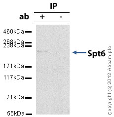
Spt6 was immunoprecipitated using 0.5mg Hela whole cell extract, 5µg of Rabbit polyclonal to Spt6 and 50µl of protein G magnetic beads (+). No antibody was added to the control (-).
The antibody was incubated under agitation with Protein G beads for 10min, Hela whole cell extract lysate diluted in RIPA buffer was added to each sample and incubated for a further 10min under agitation.
Proteins were eluted by addition of 40µl SDS loading buffer and incubated for 10min at 70oC; 10µl of each sample was separated on a SDS PAGE gel, transferred to a nitrocellulose membrane, blocked with 5% BSA and probed with ab32820.
Secondary: Mouse monoclonal [SB62a] Secondary Antibody to Rabbit IgG light chain (HRP) (ab99697).
Band: 230kDa: Spt6
Protocols
Datasheets and documents
References (9)
ab32820 has been referenced in 9 publications.
- Soboleva TA et al. A new link between transcriptional initiation and pre-mRNA splicing: The RNA binding histone variant H2A.B. PLoS Genet 13:e1006633 (2017). ChIP ; Mouse . PubMed: 28234895
- Dembowski JA et al. Replication-Coupled Recruitment of Viral and Cellular Factors to Herpes Simplex Virus Type 1 Replication Forks for the Maintenance and Expression of Viral Genomes. PLoS Pathog 13:e1006166 (2017). PubMed: 28095497
- Gérard A et al. The integrase cofactor LEDGF/p75 associates with Iws1 and Spt6 for postintegration silencing of HIV-1 gene expression in latently infected cells. Cell Host Microbe 17:107-17 (2015). PubMed: 25590759
- Dembowski JA & DeLuca NA Selective recruitment of nuclear factors to productively replicating herpes simplex virus genomes. PLoS Pathog 11:e1004939 (2015). ICC/IF . PubMed: 26018390
- Dietrich JE et al. Venus trap in the mouse embryo reveals distinct molecular dynamics underlying specification of first embryonic lineages. EMBO Rep 16:1005-21 (2015). PubMed: 26142281
- Nakamura M et al. Spt6 levels are modulated by PAAF1 and proteasome to regulate the HIV-1 LTR. Retrovirology 9:13 (2012). ICC/IF . PubMed: 22316138
- Gallastegui E et al. Chromatin Reassembly Factors Are Involved in Transcriptional Interference Promoting HIV Latency. J Virol 85:3187-202 (2011). WB . PubMed: 21270164
- Vanti M et al. Yeast genetic analysis reveals the involvement of chromatin reassembly factors in repressing HIV-1 basal transcription. PLoS Genet 5:e1000339 (2009). WB ; Human . PubMed: 19148280
- Shen X et al. Identification of novel SHPS-1-associated proteins and their roles in regulation of insulin-like growth factor-dependent responses in vascular smooth muscle cells. Mol Cell Proteomics : (2009). WB . PubMed: 19299420
Images
-
Lane 1 : Marker
Lanes 2-4 : Anti-Spt6 antibody (ab32820) at 1/500 dilution
Lane 5 : High Mark marker
Lane 2 : HeLa (Human epithelial carcinoma cell line) Whole Cell Lysate at 10 µg
Lane 3 :Jurkat whole cell lysate (ab7899) at 20 µg
Lane 4 : HeLa (Human epithelial carcinoma cell line) Nuclear Lysate at 20 µg
Secondary
Lanes 2-4 : IR Dye 680 Conjugated Goat anti Rabbit IgG (H+L) at 1/21250 dilution
Performed under reducing conditions.
Predicted band size: 199 kDa
Observed band size: 230 kDa why is the actual band size different from the predicted?
Ab32820 detects a band at approximately 230kDa. Other antibodies to this target detect a band in this region and it is understood that this band corresponds to the native Spt6 protein. -
Spt6 was immunoprecipitated using 0.5mg Hela whole cell extract, 5µg of Rabbit polyclonal to Spt6 and 50µl of protein G magnetic beads (+). No antibody was added to the control (-).
The antibody was incubated under agitation with Protein G beads for 10min, Hela whole cell extract lysate diluted in RIPA buffer was added to each sample and incubated for a further 10min under agitation.
Proteins were eluted by addition of 40µl SDS loading buffer and incubated for 10min at 70oC; 10µl of each sample was separated on a SDS PAGE gel, transferred to a nitrocellulose membrane, blocked with 5% BSA and probed with ab32820.
Secondary: Mouse monoclonal [SB62a] Secondary Antibody to Rabbit IgG light chain (HRP) (ab99697).
Band: 230kDa: Spt6


