Anti-MafB antibody (ab65953)
Key features and details
- Rabbit polyclonal to MafB
- Suitable for: WB, ICC/IF
- Reacts with: Human
- Isotype: IgG
Overview
-
Product name
Anti-MafB antibody
See all MafB primary antibodies -
Description
Rabbit polyclonal to MafB -
Host species
Rabbit -
Tested Applications & Species
See all applications and species dataApplication Species ICC/IF HumanWB Human -
Immunogen
Synthetic peptide conjugated to KLH derived from within residues 250 - 350 MafB.
Read Abcam's proprietary immunogen policy (Peptide available as ab71662.) -
Positive control
- This antibody gave a positive signal in the following lysates: HeLa Whole Cell, HepG2 Whole Cell, Caco 2 Whole Cell, Rat Thymus Tissue (data not shown), Rat Colon Tissue (data not shown)
Properties
-
Form
Liquid -
Storage instructions
Shipped at 4°C. Store at +4°C short term (1-2 weeks). Upon delivery aliquot. Store at -20°C or -80°C. Avoid freeze / thaw cycle. -
Storage buffer
pH: 7.40
Preservative: 0.02% Sodium azide
Constituent: PBS
Batches of this product that have a concentration Concentration information loading...
Purity
Immunogen affinity purifiedClonality
PolyclonalIsotype
IgGResearch areas
Associated products
-
Compatible Secondaries
-
Isotype control
-
Recombinant Protein
Applications
The Abpromise guarantee
Our Abpromise guarantee covers the use of ab65953 in the following tested applications.
The application notes include recommended starting dilutions; optimal dilutions/concentrations should be determined by the end user.
GuaranteedTested applications are guaranteed to work and covered by our Abpromise guarantee.
PredictedPredicted to work for this combination of applications and species but not guaranteed.
IncompatibleDoes not work for this combination of applications and species.
Application Species ICC/IF HumanWB HumanAll applications MouseRatChickenNon human primatesApplication Abreviews Notes WB Use a concentration of 1 µg/ml. Detects a band of approximately 38 kDa (predicted molecular weight: 36 kDa).ICC/IF Use a concentration of 5 µg/ml.Notes WB
Use a concentration of 1 µg/ml. Detects a band of approximately 38 kDa (predicted molecular weight: 36 kDa).ICC/IF
Use a concentration of 5 µg/ml.Target
-
Function
Acts as a transcriptional activator or repressor. Plays a pivotal role in regulating lineage-specific hematopoiesis by repressing ETS1-mediated transcription of erythroid-specific genes in myeloid cells. Required for monocytic, macrophage, podocyte and islet beta cell differentiation. Involved in renal tubule survival and F4/80 maturation. Activates the insulin and glucagon promoters. Together with PAX6, transactivates weakly the glucagon gene promoter through the G1 element. SUMO modification controls its transcriptional activity and ability to specify macrophage fate. Binds element G1 on the glucagon promoter (By similarity). Involved either as an oncogene or as a tumor suppressor, depending on the cell context. -
Tissue specificity
Ubiquitous. -
Involvement in disease
Defects in MAFB are the cause of multicentric carpotarsal osteolysis syndrome (MCTO) [MIM:166300]. MCTO is a rare skeletal disorder, usually presenting in early childhood with a clinical picture mimicking juvenile rheumatoid arthritis. Progressive destruction of the carpal and tarsal bone usually occurs and other bones may also be involved. Chronic renal failure is a frequent component of the syndrome. Mental retardation and minor facial anomalies have been noted in some patients. -
Sequence similarities
Belongs to the bZIP family. Maf subfamily.
Contains 1 bZIP domain. -
Domain
The leucine-zipper domain is involved in the interaction with LRPICD. -
Post-translational
modificationsPhosphorylated by GSK3 and MAPK13 on serine and threonine residues.
Sumoylated. Sumoylation on Lys-32 and Lys-297 stimulates its transcriptional repression activity and promotes macrophage differentiation from myeloid progenitors. -
Cellular localization
Nucleus. - Information by UniProt
-
Database links
- Entrez Gene: 9935 Human
- Entrez Gene: 16658 Mouse
- Entrez Gene: 54264 Rat
- GenBank: AF134157.1 Human
- Omim: 608968 Human
- SwissProt: Q9Y5Q3 Human
- SwissProt: P54841 Mouse
- SwissProt: P54842 Rat
see all -
Alternative names
- Kreisler antibody
- Kreisler (mouse) maf related leucine zipper homolog antibody
- Kreisler maf related leucine zipper homolog antibody
see all
Images
-
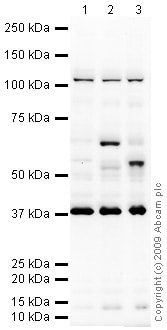
All lanes : Anti-MafB antibody (ab65953) at 1 µg/ml
Lane 1 : HeLa (Human epithelial carcinoma cell line) Whole Cell Lysate
Lane 2 : HepG2 (Human hepatocellular liver carcinoma cell line) Whole Cell Lysate
Lane 3 : Caco 2 (Human colonic carcinoma cell line) Whole Cell Lysate
Lysates/proteins at 10 µg per lane.
Secondary
All lanes : Goat polyclonal to Rabbit IgG - H&L - Pre-Adsorbed (HRP)
at 1/3000 dilution
Performed under reducing conditions.
Predicted band size: 36 kDa
Observed band size: 38 kDa why is the actual band size different from the predicted?
Additional bands at: 105 kDa, 55 kDa, 63 kDa. We are unsure as to the identity of these extra bands. -
ICC/IF image of ab65953 stained MCF-7 cells. The cells were 4% PFA fixed (10 min) and then incubated in 1%BSA / 10% normal Goat serum / 0.3M glycine in 0.1% PBS-Tween for 1h to permeabilise the cells and block non-specific protein-protein interactions. The cells were then incubated with the antibody (ab65953, 5µg/ml) overnight at +4°C. The secondary antibody (green) was Alexa Fluor® 488 Goat anti-Rabbit IgG (H+L) used at a 1/1000 dilution for 1h. Alexa Fluor® 594 WGA was used to label plasma membranes (red) at a 1/200 dilution for 1h. DAPI was used to stain the cell nuclei (blue) at a concentration of 1.43µM. This antibody also gave a positive result in 4% PFA fixed (10 min) HeLa, Hek293, HepG2 cells at 5µg/ml.
Protocols
References (2)
ab65953 has been referenced in 2 publications.
- Liu TM et al. Transcription Factor MafB Suppresses Type I Interferon Production by CD14+ Monocytes in Patients With Chronic Hepatitis C. Front Microbiol 10:1814 (2019). PubMed: 31447817
- Fujiwara T et al. RNA-binding protein Musashi2 induced by RANKL is critical for osteoclast survival. Cell Death Dis 7:e2300 (2016). WB ; Mouse . PubMed: 27441652
Images
-
All lanes : Anti-MafB antibody (ab65953) at 1 µg/ml
Lane 1 : HeLa (Human epithelial carcinoma cell line) Whole Cell Lysate
Lane 2 : HepG2 (Human hepatocellular liver carcinoma cell line) Whole Cell Lysate
Lane 3 : Caco 2 (Human colonic carcinoma cell line) Whole Cell Lysate
Lysates/proteins at 10 µg per lane.
Secondary
All lanes : Goat polyclonal to Rabbit IgG - H&L - Pre-Adsorbed (HRP)
at 1/3000 dilution
Performed under reducing conditions.
Predicted band size: 36 kDa
Observed band size: 38 kDa why is the actual band size different from the predicted?
Additional bands at: 105 kDa, 55 kDa, 63 kDa. We are unsure as to the identity of these extra bands.
-
ICC/IF image of ab65953 stained MCF-7 cells. The cells were 4% PFA fixed (10 min) and then incubated in 1%BSA / 10% normal Goat serum / 0.3M glycine in 0.1% PBS-Tween for 1h to permeabilise the cells and block non-specific protein-protein interactions. The cells were then incubated with the antibody (ab65953, 5µg/ml) overnight at +4°C. The secondary antibody (green) was Alexa Fluor® 488 Goat anti-Rabbit IgG (H+L) used at a 1/1000 dilution for 1h. Alexa Fluor® 594 WGA was used to label plasma membranes (red) at a 1/200 dilution for 1h. DAPI was used to stain the cell nuclei (blue) at a concentration of 1.43µM. This antibody also gave a positive result in 4% PFA fixed (10 min) HeLa, Hek293, HepG2 cells at 5µg/ml.


