Anti-Loricrin antibody (ab85679)
")
Key features and details
- Rabbit polyclonal to Loricrin
- Suitable for: IHC-P, ICC/IF
- Reacts with: Mouse, Human
- Isotype: IgG
Overview
-
Product name
Anti-Loricrin antibody
See all Loricrin primary antibodies -
Description
Rabbit polyclonal to Loricrin -
Host species
Rabbit -
Tested Applications & Species
See all applications and species dataApplication Species ICC/IF HumanIHC-P MouseHuman
-
Immunogen
Synthetic peptide corresponding to Mouse Loricrin aa 450 to the C-terminus (C terminal) conjugated to keyhole limpet haemocyanin.
(Peptide available asab95418) -
Positive control
- This antibody gave a positive signal in T47D (Human breast duct epithelial carcinoma) whole cell lysate. IHC-P: FFPE human and mouse normal skin tissue sections.
-
General notes
The Life Science industry has been in the grips of a reproducibility crisis for a number of years. Abcam is leading the way in addressing the problem with our range of recombinant monoclonal antibodies and knockout edited cell lines for gold-standard validation.
One factor contributing to the crisis is the use of antibodies that are not suitable. This can lead to misleading results and the use of incorrect data informing project assumptions and direction. To help address this challenge, we have introduced an application and species grid on our primary antibody datasheets to make it easy to simplify identification of the right antibody for your needs.
Learn more here.
Properties
-
Form
Liquid -
Storage instructions
Shipped at 4°C. Store at +4°C short term (1-2 weeks). Upon delivery aliquot. Store at -20°C or -80°C. Avoid freeze / thaw cycle. -
Storage buffer
pH: 7.40
Preservative: 0.02% Sodium azide
Constituent: PBS
Batches of this product that have a concentration Concentration information loading...
Purity
Immunogen affinity purifiedClonality
PolyclonalIsotype
IgGResearch areas
Associated products
-
Compatible Secondaries
-
Immunizing Peptide (Blocking)
-
Isotype control
-
Recombinant Protein
Applications
The Abpromise guarantee
Our Abpromise guarantee covers the use of ab85679 in the following tested applications.
The application notes include recommended starting dilutions; optimal dilutions/concentrations should be determined by the end user.
GuaranteedTested applications are guaranteed to work and covered by our Abpromise guarantee.
PredictedPredicted to work for this combination of applications and species but not guaranteed.
IncompatibleDoes not work for this combination of applications and species.
Application Species ICC/IF HumanIHC-P MouseHumanAll applications RatApplication Abreviews Notes IHC-P (5) Use a concentration of 5 µg/ml.ICC/IF (1) Use a concentration of 5 µg/ml.Notes IHC-P
Use a concentration of 5 µg/ml.ICC/IF
Use a concentration of 5 µg/ml.Target
-
Function
Major keratinocyte cell envelope protein. -
Involvement in disease
Defects in LOR are a cause of progressive symmetric erythrokeratodermia (PSEK) [MIM:133200]. Erythrokeratodermas are a group of disorders characterized by widespread erythematous plaques, either stationary or migratory, associated with features that include palmoplantar keratoderma. PSEK is characterized by erythematous and hyperkeratotic plaques.
Defects in LOR are the cause of Vohwinkel syndrome with ichthyosis (VSI) [MIM:604117]; also known as loricrin keratoderma (LK) or mutilating keratoderma with ichthyosis. VSI is an ichthyotic variant of Vohwinkel syndrome (VS) characterized by progressive symmetric erythrokeratoderma or congenital ichthyosiform erythroderma born as a collodion baby. Common clinical features include hyperkeratosis of the palms and soles with digital constriction. -
Post-translational
modificationsSubstrate of transglutaminases. Some glutamines and lysines are cross-linked to other loricrin molecules and to SPRRs proteins.
Contains inter- or intramolecular disulfide-bonds. -
Cellular localization
Cytoplasm. Nucleus > nucleoplasm. - Information by UniProt
-
Database links
- Entrez Gene: 4014 Human
- Entrez Gene: 16939 Mouse
- Entrez Gene: 502541 Rat
- Omim: 152445 Human
- SwissProt: P23490 Human
- SwissProt: P18165 Mouse
- Unigene: 251680 Human
- Unigene: 1121 Mouse
-
Alternative names
- LOR antibody
- LOR protein antibody
- LORI_HUMAN antibody
see all
Images
-
Immunohistochemistry (Formalin/PFA-fixed paraffin-embedded sections) - Anti-Loricrin antibody (ab85679)
IHC image of Loricrin staining in Mouse normal skin formalin fixed paraffin embedded tissue section, performed on a Leica Bond™ system using the standard protocol B. The section was pre-treated using heat mediated antigen retrieval with sodium citrate buffer (pH6, epitope retrieval solution 1) for 20 mins. The section was then incubated with ab85679, 5µg/ml, for 15 mins at room temperature and detected using an HRP conjugated compact polymer system. DAB was used as the chromogen. The section was then counterstained with haematoxylin and mounted with DPX.
For other IHC staining systems (automated and non-automated) customers should optimize variable parameters such as antigen retrieval conditions, primary antibody concentration and antibody incubation times.
-
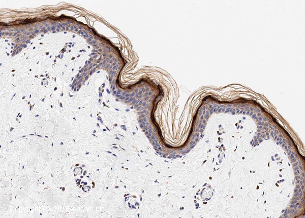 Immunohistochemistry (Formalin/PFA-fixed paraffin-embedded sections) - Anti-Loricrin antibody (ab85679)
IHC image of Loricrin staining in human normal skin formalin fixed paraffin embedded tissue section*, performed on a Leica Bond™ system using the standard protocol F. The section was pre-treated using heat mediated antigen retrieval with sodium citrate buffer (pH6, epitope retrieval solution 1) for 20 mins. The section was then incubated with ab85679, 1µg/ml, for 15 mins at room temperature and detected using an HRP conjugated compact polymer system. DAB was used as the chromogen. The section was then counterstained with haematoxylin and mounted with DPX.
For other IHC staining systems (automated and non-automated) customers should optimize variable parameters such as antigen retrieval conditions, primary antibody concentration and antibody incubation times.
*Tissue obtained from the Human Research Tissue Bank, supported by the NIHR Cambridge Biomedical Research Centre
-
 Immunocytochemistry/ Immunofluorescence - Anti-Loricrin antibody (ab85679)ICC/IF image of ab85679 stained HepG2 cells. The cells were 4% PFA fixed (10 min) and then incubated in 1%BSA / 10% normal Goat serum / 0.3M glycine in 0.1% PBS-Tween for 1h to permeabilise the cells and block non-specific protein-protein interactions. The cells were then incubated with the antibody (ab85679, 5µg/ml) overnight at +4°C. The secondary antibody (green) was Alexa Fluor® 488 Goat anti-Rabbit IgG (H+L) used at a 1/1000 dilution for 1h. Alexa Fluor® 594 WGA was used to label plasma membranes (red) at a 1/200 dilution for 1h. DAPI was used to stain the cell nuclei (blue) at a concentration of 1.43µM.
Protocols
Datasheets and documents
-
SDS download
-
Datasheet download
References (18)
ab85679 has been referenced in 18 publications.
- Hieda DS et al. Air Particulate Matter Induces Skin Barrier Dysfunction and Water Transport Alteration on a Reconstructed Human Epidermis Model. J Invest Dermatol N/A:N/A (2020). PubMed: 32339540
- Zhang Q et al. Early-stage bilayer tissue-engineered skin substitute formed by adult skin progenitor cells produces an improved skin structure in vivo. Stem Cell Res Ther 11:407 (2020). PubMed: 32948249
- Welz PS et al. BMAL1-Driven Tissue Clocks Respond Independently to Light to Maintain Homeostasis. Cell 177:1436-1447.e12 (2019). PubMed: 31150620
- Todorovic V et al. Small Molecule IL-36? Antagonist as a Novel Therapeutic Approach for Plaque Psoriasis. Sci Rep 9:9089 (2019). PubMed: 31235749
- Nasir NAM et al. Fluorescent cell tracer dye permits real-time assessment of re-epithelialization in a serum-free ex vivo human skin wound assay. Wound Repair Regen 27:126-133 (2019). PubMed: 30575205
Images
-
Immunohistochemistry (Formalin/PFA-fixed paraffin-embedded sections) - Anti-Loricrin antibody (ab85679)
IHC image of Loricrin staining in Mouse normal skin formalin fixed paraffin embedded tissue section, performed on a Leica Bond™ system using the standard protocol B. The section was pre-treated using heat mediated antigen retrieval with sodium citrate buffer (pH6, epitope retrieval solution 1) for 20 mins. The section was then incubated with ab85679, 5µg/ml, for 15 mins at room temperature and detected using an HRP conjugated compact polymer system. DAB was used as the chromogen. The section was then counterstained with haematoxylin and mounted with DPX.
For other IHC staining systems (automated and non-automated) customers should optimize variable parameters such as antigen retrieval conditions, primary antibody concentration and antibody incubation times.
-
Immunohistochemistry (Formalin/PFA-fixed paraffin-embedded sections) - Anti-Loricrin antibody (ab85679)
IHC image of Loricrin staining in human normal skin formalin fixed paraffin embedded tissue section*, performed on a Leica Bond™ system using the standard protocol F. The section was pre-treated using heat mediated antigen retrieval with sodium citrate buffer (pH6, epitope retrieval solution 1) for 20 mins. The section was then incubated with ab85679, 1µg/ml, for 15 mins at room temperature and detected using an HRP conjugated compact polymer system. DAB was used as the chromogen. The section was then counterstained with haematoxylin and mounted with DPX.
For other IHC staining systems (automated and non-automated) customers should optimize variable parameters such as antigen retrieval conditions, primary antibody concentration and antibody incubation times.
*Tissue obtained from the Human Research Tissue Bank, supported by the NIHR Cambridge Biomedical Research Centre
-
Immunocytochemistry/ Immunofluorescence - Anti-Loricrin antibody (ab85679)ICC/IF image of ab85679 stained HepG2 cells. The cells were 4% PFA fixed (10 min) and then incubated in 1%BSA / 10% normal Goat serum / 0.3M glycine in 0.1% PBS-Tween for 1h to permeabilise the cells and block non-specific protein-protein interactions. The cells were then incubated with the antibody (ab85679, 5µg/ml) overnight at +4°C. The secondary antibody (green) was Alexa Fluor® 488 Goat anti-Rabbit IgG (H+L) used at a 1/1000 dilution for 1h. Alexa Fluor® 594 WGA was used to label plasma membranes (red) at a 1/200 dilution for 1h. DAPI was used to stain the cell nuclei (blue) at a concentration of 1.43µM.


