Anti-Lamin A antibody (ab26300)
")
Key features and details
- Rabbit polyclonal to Lamin A
- Suitable for: ICC/IF, WB
- Knockout validated
- Reacts with: Mouse, Rat, Human
- Isotype: IgG
Overview
-
Product name
Anti-Lamin A antibody
See all Lamin A primary antibodies -
Description
Rabbit polyclonal to Lamin A -
Host species
Rabbit -
Tested Applications & Species
See all applications and species dataApplication Species ICC/IF HumanWB RatHuman
-
Immunogen
Synthetic peptide conjugated to KLH derived from within residues 550 to the C-terminus of Human Lamin A.
Read Abcam's proprietary immunogen policy (Peptide available as ab27812.) -
Positive control
- ab26300 gave a positive result in the following Whole Cell Lysates A431 NIH 3T3 PC12 ICC-IF: Hela cells
Images
-
Western blot - Anti-Lamin A antibody (ab26300)
Lane 1: Wild-type HAP1 cell lysate (20 µg)
Lane 2: Lamin A knockout HAP1 cell lysate (20 µg)
Lane 3: A431 cell lysate (20 µg)
Lane 4: NIH3T3 cell lysate (20 µg)
Lanes 1 - 4: Merged signal (red and green). Green - ab26300 observed at 76 kDa. Red - loading control, ab8245, observed at 37 kDa.
ab26300 was shown to recognize Lamin A in wild-type HAP1 cells along with additional cross-reactive bands. No band was observed when Lamin A knockout samples were examined. Wild-type and Lamin A knockout samples were subjected to SDS-PAGE. ab26300 1ug/ml and ab8245 (loading control to GAPDH) at a dilution of 1/1000 were incubated overnight at 4°C. Blots were developed with Goat anti-Rabbit IgG H&L (IRDye® 800CW) preadsorbed (ab216773) and Goat anti-Mouse IgG H&L (IRDye® 680RD) preadsorbed (ab216776) secondary antibodies at 1/10,000 dilution for 1 hour at room temperature before imaging. -
 Immunocytochemistry/ Immunofluorescence - Anti-Lamin A antibody (ab26300)
ab26300 stained in Hela cells. Cells were fixed with 100% methanol (5 min) at room temperature and incubated with PBS containing 10% goat serum, 0.3 M glycine, 1% BSA and 0.1% triton for 1h at room temperature to permeabilise the cells and block non-specific protein-protein interactions. The cells were then incubated with the antibody ab26300 at 1µg/ml and ab7291 (Mouse monoclonal [DM1A] to alpha Tubulin - Loading Control) at 1/1000 dilution overnight at +4°C. The secondary antibodies were ab150120 (pseudo-colored red) and ab150081 (colored green) used at 1 ug/ml for 1hour at room temperature. DAPI was used to stain the cell nuclei (colored blue) at a concentration of 1.43µM for 1hour at room temperature.
-
 Immunocytochemistry/ Immunofluorescence - Anti-Lamin A antibody (ab26300) Image from Khatau, Shyam B. et al. PLoS ONE 7.5 (2012): e36689. doi: 10.1371/journal.pone.0036689. Fig 5A. Reproduced under the Creative Commons license https://creativecommons.org/publicdomain/zero/1.0/
Immunocytochemistry/ Immunofluorescence analysis of hESCs labeling Lamin A with ab26300 at 1/500 dilution. Samples were fixed with 3.7% paraformaldehyde for 1 hour, and stained for nuclear DNA (DAPI), filamentous actin, tumor recognition antigen 1–81, and nuclear envelope protein Lamin A. For staining, cells were permeabilized with 0.1% Triton X-100 for 10 min. Goat serum, 10%, in phosphate-buffered saline was used to block nonspecific binding for 20 min.
-
 Immunocytochemistry/ Immunofluorescence - Anti-Lamin A antibody (ab26300) This image is part of an Abreview submmited by Dr Kirk McManus.ab26300 (1/2000) staining Lamin A in assynchronous HeLa Cells, by Immunocytochemistry/ Immunofluorescence. Secondary antibody: goat anti-rabbit conjugated to Cy3 ® (1/200). Cells counterstained with DAPI in order to highlight the nucleus.
 -
 Western blot - Anti-Lamin A antibody (ab26300) This image is courtesy of an anonymous abreview.Anti-Lamin A antibody (ab26300) at 1/1000 dilution + HeLa whole cell extract at 100 µg
Secondary
Goat anti-Rabbit IgG (H+L) HRP Conjugate at 1/10000 dilution
Developed using the ECL technique.
Predicted band size: 74 kDa
Observed band size: 76 kDa why is the actual band size different from the predicted?
Exposure time: 15 secondsBlocking: 5% milk for 30 minutes at 22°C
-
 Immunocytochemistry/ Immunofluorescence - Anti-Lamin A antibody (ab26300) This image is courtesy of an anonymous abreview.
Immunocytochemistry/ Immunofluorescence analysis of human vascular smooth muscle cell labeling Lamin A with ab26300 at 1/200 dilution. Cells were fixed in formaldehyde and permeabilized with np40. Cells were blocked with 3% BSA for 1 hour at 21°C. A polyclonal donkey anti-rabbit Alexa Fluor® 568 conjugated secondary antibody was used at 1/500 dilution.
-
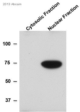 Western blot - Anti-Lamin A antibody (ab26300) This image is courtesy of an anonymous AbreviewAll lanes : Anti-Lamin A antibody (ab26300) at 1/1000 dilution
Lane 1 : Mouse NIH-3T3 cells - cytosolic fraction
Lane 2 : Mouse NIH-3T3 cells - nuclear fraction
Lysates/proteins at 25 µg per lane.
Secondary
All lanes : HRP conjugated Goat anti-rabbit at 1/5000 dilution
Developed using the ECL technique.
Performed under reducing conditions.
Predicted band size: 74 kDa
Observed band size: 76 kDa why is the actual band size different from the predicted?
Exposure time: 10 seconds
-
 Immunocytochemistry/ Immunofluorescence - Anti-Lamin A antibody (ab26300) Image from Khatau, Shyam B. et al. PLoS ONE 7.5 (2012): e36689. doi: 10.1371/journal.pone.0036689. Fig 5E.
Immunocytochemistry/ Immunofluorescence analysis of Human Lung Fibroblasts labeling Lamin A with ab26300 at 1/500 dilution. Samples were fixed with 3.7% paraformaldehyde for 1 hour, and stained for nuclear DNA (DAPI), filamentous actin, tumor recognition antigen 1–81, and nuclear envelope protein Lamin A. For staining, cells were permeabilized with 0.1% Triton X-100 for 10 min. Goat serum, 10%, in phosphate-buffered saline was used to block nonspecific binding for 20 min.
-
 Western blot - Anti-Lamin A antibody (ab26300)Anti-Lamin A antibody (ab26300) at 1 µg/ml +
A-431 whole cell lysate (ab7909) at 20 µg
Secondary
IRDye 680 Conjugated Goat Anti-Rabbit IgG (H+L) at 1/15000 dilution
Performed under reducing conditions.
Predicted band size: 74 kDa
Observed band size: 76 kDa why is the actual band size different from the predicted?
Additional bands at: 68 kDa (possible degradation product) -
 Western blot - Anti-Lamin A antibody (ab26300)All lanes : Anti-Lamin A antibody (ab26300) at 1 µg/ml
Lane 1 :Recombinant Human Lamin A protein (ab83472) at 0.1 µg
Lane 2 :Recombinant Human Lamin A protein (ab83472) at 0.01 µg
Secondary
All lanes : Goat Anti-Rabbit IgG H&L (HRP) preadsorbed (ab97080) at 1/5000 dilution
Developed using the ECL technique.
Performed under reducing conditions.
Predicted band size: 74 kDa
Exposure time: 10 seconds -
 Immunocytochemistry/ Immunofluorescence - Anti-Lamin A antibody (ab26300) This image is courtesy of an Abreview submitted by Dr Chi W Tangab26300 at 1/1000 staining human HeLa cells by ICC/IF. The cells were paraformaldehyde fixed, permeabilized with Triton X100 and blocked with BSA before incubation with the antibody. A Cy3 ® conjugated donkey anti-rabbit IgG was used as the secondary.
 -
 Western blot - Anti-Lamin A antibody (ab26300)All lanes : Anti-Lamin A antibody (ab26300) at 1 µg/ml
Lane 1 :NIH/3T3 whole cell lysate (ab7179)
Lane 2 : PC12 (Rat adrenal pheochromocytoma cell line) Whole Cell Lysate
Lysates/proteins at 10 µg per lane.
Secondary
All lanes : IRDye 680 Conjugated Goat Anti-Rabbit IgG (H+L) at 1/10000 dilution
Performed under reducing conditions.
Predicted band size: 74 kDa
Observed band size: 74 kDa
Additional bands at: 100 kDa, 45 kDa, 70 kDa (possible degradation product). We are unsure as to the identity of these extra bands.


