Anti-Kindlin-1 antibody (ab68041)
Key features and details
- Rabbit polyclonal to Kindlin-1
- Suitable for: ICC/IF, IHC-P, WB
- Reacts with: Mouse, Human
- Isotype: IgG
Overview
-
Product name
Anti-Kindlin-1 antibody -
Description
Rabbit polyclonal to Kindlin-1 -
Host species
Rabbit -
Tested applications
Suitable for: ICC/IF, IHC-P, WBmore details -
Species reactivity
Reacts with: Mouse, Human
Predicted to work with: Cow, Xenopus laevis, Non human primates
-
Immunogen
-
General notes
This product was previously labelled as Kindlin
Reproducibility is key to advancing scientific discovery and accelerating scientists’ next breakthrough.
Abcam is leading the way with our range of recombinant antibodies, knockout-validated antibodies and knockout cell lines, all of which support improved reproducibility.
We are also planning to innovate the way in which we present recommended applications and species on our product datasheets, so that only applications & species that have been tested in our own labs, our suppliers or by selected trusted collaborators are covered by our Abpromise™ guarantee.
In preparation for this, we have started to update the applications & species that this product is Abpromise guaranteed for.
We are also updating the applications & species that this product has been “predicted to work with,” however this information is not covered by our Abpromise guarantee.
Applications & species from publications and Abreviews that have not been tested in our own labs or in those of our suppliers are not covered by the Abpromise guarantee.
Please check that this product meets your needs before purchasing. If you have any questions, special requirements or concerns, please send us an inquiry and/or contact our Support team ahead of purchase. Recommended alternatives for this product can be found below, as well as customer reviews and Q&As.
Properties
-
Form
Liquid -
Storage instructions
Shipped at 4°C. Store at +4°C short term (1-2 weeks). Upon delivery aliquot. Store at -20°C or -80°C. Avoid freeze / thaw cycle. -
Storage buffer
pH: 7.40
Preservative: 0.02% Sodium azide
Constituent: PBS
Batches of this product that have a concentration Concentration information loading...
Purity
Immunogen affinity purifiedClonality
PolyclonalIsotype
IgGResearch areas
Associated products
-
Compatible Secondaries
-
Immunizing Peptide (Blocking)
-
Isotype control
-
Recombinant Protein
Applications
Our Abpromise guarantee covers the use of ab68041 in the following tested applications.
The application notes include recommended starting dilutions; optimal dilutions/concentrations should be determined by the end user.
Application Abreviews Notes ICC/IF Use a concentration of 5 µg/ml. IHC-P Use a concentration of 5 µg/ml. Perform heat mediated antigen retrieval before commencing with IHC staining protocol. WB Use a concentration of 1 µg/ml. Detects a band of approximately 77 kDa (predicted molecular weight: 77 kDa). Target
-
Function
Involved in cell adhesion. Contributes to integrin activation. When coexpressed with talin, potentiates activation of ITGA2B. Required for normal keratinocyte proliferation. Required for normal polarization of basal keratinocytes in skin, and for normal cell shape. Required for normal adhesion of keratinocytes to fibronectin and laminin, and for normal keratinocyte migration to wound sites. May mediate TGF-beta 1 signaling in tumor progression. -
Tissue specificity
Expressed in brain, skeletal muscle, kidney, colon, adrenal gland, prostate, and placenta. Weakly or not expressed in heart, thymus, spleen, liver, small intestine, bone marrow, lung and peripheral blood leukocytes. Overexpressed in some colon and lung tumors. In skin, it is localized within the epidermis and particularly in basal keratocytes. Not detected in epidermal melanocytes and dermal fibroblasts. -
Involvement in disease
Defects in FERMT1 are the cause of Kindler syndrome (KINDS) [MIM:173650]. An autosomal recessive skin disorder characterized by skin blistering, photosensitivity, progressive poikiloderma, and extensive skin atrophy. Additional clinical features include gingival erosions, ocular, esophageal, gastrointestinal and urogenital involvement, and an increased risk of mucocutaneous malignancy. Note=Although most FERMT1 mutations are predicted to lead to premature termination of translation, and to loss of FERMT1 function, significant clinical variability is observed among patients. There is an association of FERMT1 missense and in-frame deletion mutations with milder disease phenotypes, and later onset of complications (PubMed:21936020). -
Sequence similarities
Belongs to the kindlin family.
Contains 1 FERM domain.
Contains 1 PH domain. -
Domain
The FERM domain is not correctly detected by PROSITE or Pfam techniques because it contains the insertion of a PH domain. The FERM domain contains the subdomains F1, F2 and F3. It is preceded by a F0 domain with a ubiquitin-like fold. The F0 domain is required for integrin activation and for localization at focal adhesions. -
Cellular localization
Cytoplasm > cytoskeleton. Cell junction > focal adhesion. Cell projection > ruffle membrane. Constituent of focal adhesions. Localized at the basal aspect of skin keratinocytes, close to the cell membrane. Colocalizes with filamentous actin. Upon TGFB1 treatment, it localizes to membrane ruffles. - Information by UniProt
-
Database links
- Entrez Gene: 524427 Cow
- Entrez Gene: 55612 Human
- Entrez Gene: 241639 Mouse
- Omim: 607900 Human
- SwissProt: Q9BQL6 Human
- SwissProt: P59113 Mouse
- Unigene: 472054 Human
- Unigene: 209784 Mouse
-
Alternative names
- C20orf42 antibody
- Chromosome 20 open reading frame 42 antibody
- DTGCU 2 antibody
see all
Images
-
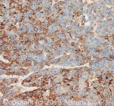
 Immunohistochemistry (Formalin/PFA-fixed paraffin-embedded sections) - Anti-Kindlin-1 antibody (ab68041)
IHC image of Kindlin-1 staining in human liver carcnioma formalin fixed paraffin embedded tissue section, performed on a Leica BondTM system using the standard protocol F. The section was pre-treated using heat mediated antigen retrieval with sodium citrate buffer (pH6, epitope retrieval solution 1) for 20 mins. The section was then incubated with ab68041, 5µg/ml, for 15 mins at room temperature and detected using an HRP conjugated compact polymer system. DAB was used as the chromogen. The section was then counterstained with haematoxylin and mounted with DPX.
For other IHC staining systems (automated and non-automated) customers should optimize variable parameters such as antigen retrieval conditions, primary antibody concentration and antibody incubation times. -
All lanes : Anti-Kindlin-1 antibody (ab68041) at 1 µg/ml
Lane 1 : Jurkat Whole Cell Lysate - Staurosporine Treated (24hr, 500nM)
Lane 2 : TE 671 (Human Rhabdomyosarcoma) Whole Cell Lysate
Lane 3 : Y79 (Human retinoblastoma cell line) Whole Cell Lysate
Lane 4 : MEF1 (Mouse embryonic fibroblast cell line) Whole Cell Lysate
Lane 5 : Hela Whole Cell Lysate - Staurosporine Treated (24hr, 500nM)
Lysates/proteins at 10 µg per lane.
Secondary
All lanes : Goat polyclonal to Rabbit IgG - H&L - Pre-Adsorbed (HRP)
Performed under reducing conditions.
Predicted band size: 77 kDa
Observed band size: 77 kDa
Additional bands at: 45 kDa, 74 kDa. We are unsure as to the identity of these extra bands. -
ICC/IF image of ab68041 stained HeLa cells. The cells were 4% PFA fixed (10 min) and then incubated in 1%BSA / 10% normal goat serum / 0.3M glycine in 0.1% PBS-Tween for 1h to permeabilise the cells and block non-specific protein-protein interactions. The cells were then incubated with the antibody (ab68041, 5µg/ml) overnight at +4°C. The secondary antibody (green) was Alexa Fluor® 488 goat anti-rabbit IgG (H+L) used at a 1/1000 dilution for 1h. Alexa Fluor® 594 WGA was used to label plasma membranes (red) at a 1/200 dilution for 1h. DAPI was used to stain the cell nuclei (blue) at a concentration of 1.43µM.
Protocols
References (10)
ab68041 has been referenced in 10 publications.
- Rostampour N et al. Expression of new genes in vertebrate tooth development and p63 signaling. Dev Dyn N/A:N/A (2019). PubMed: 30875130
- Azorin P et al. Distinct expression profiles and functions of Kindlins in breast cancer. J Exp Clin Cancer Res 37:281 (2018). PubMed: 30477537
- Georgiadou M et al. AMPK negatively regulates tensin-dependent integrin activity. J Cell Biol 216:1107-1121 (2017). PubMed: 28289092
- Patel H et al. Kindlin1 regulates microtubule function to ensure normal mitosis. J Mol Cell Biol 8:338-48 (2016). WB ; Human . PubMed: 26993041
- Inaba J et al. Lunasin sensitivity in non-small cell lung cancer cells is linked to suppression of integrin signaling and changes in histone acetylation. Int J Mol Sci 15:23705-24 (2014). ICC/IF ; Human . PubMed: 25530619
- Mahawithitwong P et al. Kindlin-1 expression is involved in migration and invasion of pancreatic cancer. Int J Oncol 42:1360-6 (2013). PubMed: 23440354
- Rantala JK et al. SHARPIN is an endogenous inhibitor of ß1-integrin activation. Nat Cell Biol 13:1315-24 (2011). PubMed: 21947080
- Natsuga K et al. Expression of exon-8-skipped kindlin-1 does not compensate for defects of Kindler syndrome. J Dermatol Sci 61:38-44 (2011). WB, ICC/IF ; Human . PubMed: 21146372
- Bauer R et al. Inhibition of collagen XVI expression reduces glioma cell invasiveness. Cell Physiol Biochem 27:217-26 (2011). WB ; Human . PubMed: 21471710
- Nevo J et al. Mammary-derived growth inhibitor (MDGI) interacts with integrin a-subunits and suppresses integrin activity and invasion. Oncogene 29:6452-63 (2010). ICC/IF ; Human . PubMed: 20802519
Images
-
 Immunohistochemistry (Formalin/PFA-fixed paraffin-embedded sections) - Anti-Kindlin-1 antibody (ab68041)
IHC image of Kindlin-1 staining in human liver carcnioma formalin fixed paraffin embedded tissue section, performed on a Leica BondTM system using the standard protocol F. The section was pre-treated using heat mediated antigen retrieval with sodium citrate buffer (pH6, epitope retrieval solution 1) for 20 mins. The section was then incubated with ab68041, 5µg/ml, for 15 mins at room temperature and detected using an HRP conjugated compact polymer system. DAB was used as the chromogen. The section was then counterstained with haematoxylin and mounted with DPX.
For other IHC staining systems (automated and non-automated) customers should optimize variable parameters such as antigen retrieval conditions, primary antibody concentration and antibody incubation times. -
All lanes : Anti-Kindlin-1 antibody (ab68041) at 1 µg/ml
Lane 1 : Jurkat Whole Cell Lysate - Staurosporine Treated (24hr, 500nM)
Lane 2 : TE 671 (Human Rhabdomyosarcoma) Whole Cell Lysate
Lane 3 : Y79 (Human retinoblastoma cell line) Whole Cell Lysate
Lane 4 : MEF1 (Mouse embryonic fibroblast cell line) Whole Cell Lysate
Lane 5 : Hela Whole Cell Lysate - Staurosporine Treated (24hr, 500nM)
Lysates/proteins at 10 µg per lane.
Secondary
All lanes : Goat polyclonal to Rabbit IgG - H&L - Pre-Adsorbed (HRP)
Performed under reducing conditions.
Predicted band size: 77 kDa
Observed band size: 77 kDa
Additional bands at: 45 kDa, 74 kDa. We are unsure as to the identity of these extra bands.
-
ICC/IF image of ab68041 stained HeLa cells. The cells were 4% PFA fixed (10 min) and then incubated in 1%BSA / 10% normal goat serum / 0.3M glycine in 0.1% PBS-Tween for 1h to permeabilise the cells and block non-specific protein-protein interactions. The cells were then incubated with the antibody (ab68041, 5µg/ml) overnight at +4°C. The secondary antibody (green) was Alexa Fluor® 488 goat anti-rabbit IgG (H+L) used at a 1/1000 dilution for 1h. Alexa Fluor® 594 WGA was used to label plasma membranes (red) at a 1/200 dilution for 1h. DAPI was used to stain the cell nuclei (blue) at a concentration of 1.43µM.


