Anti-KDM1/LSD1 antibody - Nuclear Marker (ab17721)
Key features and details
- Rabbit polyclonal to KDM1/LSD1 - Nuclear Marker
- Suitable for: ICC/IF, WB
- Knockout validated
- Reacts with: Mouse, Rat, Human
- Isotype: IgG
Overview
-
Product name
Anti-KDM1/LSD1 antibody - Nuclear Marker
See all KDM1/LSD1 primary antibodies -
Description
Rabbit polyclonal to KDM1/LSD1 - Nuclear Marker -
Host species
Rabbit -
Tested Applications & Species
See all applications and species dataApplication Species ICC/IF HumanWB MouseRatHuman -
Immunogen
Synthetic peptide conjugated to KLH derived from within residues 800 to the C-terminus of Human LSD1.
Read Abcam's proprietary immunogen policy (Peptide available as ab17763.)
Images
-
Lane 1: Wild type HAP1 whole cell lysate (20 µg)
Lane 2: KDM1 / LSD1 knockout HAP1 whole cell lysate (20 µg)
Lane 3: HeLa whole cell lysate (20 µg)
Lane 4: Jurkat whole cell lysate (20 µg)
Lanes 1 - 4: Merged signal (red and green). Green - ab17721 observed at 110 kDa. Red - loading control, ab8245, observed at 37 kDa.ab17721 was shown to specifically react with KDMI/LSD1 in wild-type HAP1 cells. No band was observed when KDMI/LSD1 knockout samples were examined.
-
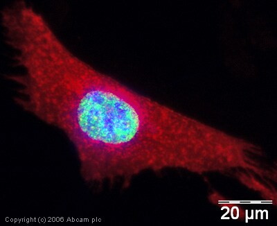
 Immunocytochemistry/ Immunofluorescence - Anti-KDM1/LSD1 antibody - Nuclear Marker and ChIP Grade (ab17721)
ICC/IF image of ab17721 stained human HeLa cells. The cells were methanol fixed (5 min) and incubated with the antibody (ab17721, 1µg/ml) for 1h at room temperature. The secondary antibody (green) was Alexa Fluor® 488 goat anti-rabbit IgG (H+L) used at a 1/1000 dilution for 1h. Image-iTTM FX Signal Enhancer was used as the primary blocking agent, 5% BSA (in TBS-T) was used for all other blocking steps. DAPI was used to stain the cell nuclei (blue). Alexa Fluor® 594 WGA was used to label plasma membranes (red).
-
All lanes :
Lane 1 : Jurkat (Human T cell leukemia T lymphocyte) whole cell lysates at 20 µg
Lane 2 : HCT 116 (Human colorectal carcinoma epithelial cell) whole cell lysates at 20 µg
Lane 3 : NIH/3T3 (Mouse embryonic fibroblast) whole cell lysates at 20 µg
Lane 4 : C2C12 (Mouse myoblasts myoblast) whole cell lysates
Lane 5 : Mouse skeletal muscle lysates at 20 µg
Lane 6 : C6 (Rat glial tumor glial cell) whole cell lysates at 20 µg
Secondary
All lanes : Goat Anti-Rabbit IgG, (H+L), Peroxidase conjugated (ab97051) at 1/20000 dilution
Predicted band size: 93 kDa
Exposure time: 50 secondsObserved MW: 110kd
Blocking Buffer & Diluting buffer concentration: 5% NFDM/TBST
-
All lanes : Anti-KDM1/LSD1 antibody - Nuclear Marker (ab17721) at 1 µg/ml
Lane 1 : HeLa (Human epithelial carcinoma cell line) Whole Cell Lysate
Lane 2 :Jurkat whole cell lysate (ab7899)
Lane 3 :NIH/3T3 whole cell lysate (ab7179)
Lysates/proteins at 20 µg per lane.
Secondary
All lanes : Goat polyclonal to Rabbit IgG H&L (HRP) Pre-Adsorbed at 1/10000 dilution
Performed under reducing conditions.
Predicted band size: 93 kDa -
 Immunocytochemistry/ Immunofluorescence - Anti-KDM1/LSD1 antibody - Nuclear Marker and ChIP Grade (ab17721) Image courtesy of anonymous abreviewer
ab17721 staining LSD1 in the mouse c2c12 cell line by ICC/IF (Immunocytochemistry/immunofluorescence). Cells were fixed with paraformaldehyde, permeabilized with 0.5% Triton X-100 and blocked with 2% BSA for 1 hour at 25°C. Samples were incubated with primary antibody (1/200) for 1 hour at 25°C. A diluted (1/2000) Alexa Fluor® 568-conjugated Goat anti-rabbit IgG polyclonal was used as the secondary antibody.
-
 Immunocytochemistry/ Immunofluorescence - Anti-KDM1/LSD1 antibody - Nuclear Marker and ChIP Grade (ab17721) This image is courtesy of Katia AncelinHuman 293T cells fixed in 4% paraformaldehyde were immunostained with ab17721 (1/200) for LSD1 and detected using FITC labelled anti-rabbit (Green). Nuclear DNA is stained blue with DAPI.


