Anti-JDP2 antibody (ab40916)
Key features and details
- Rabbit polyclonal to JDP2
- Suitable for: IHC-P, WB, ICC/IF
- Reacts with: Human
- Isotype: IgG
Overview
-
Product name
Anti-JDP2 antibody -
Description
Rabbit polyclonal to JDP2 -
Host species
Rabbit -
Tested applications
Suitable for: IHC-P, WB, ICC/IFmore details -
Species reactivity
Reacts with: Human
Predicted to work with: Rat Does not react with:
Mouse
 -
Immunogen
Synthetic peptide conjugated to KLH derived from within residues 100 to the C-terminus of Human JDP2.
Read Abcam's proprietary immunogen policy (Peptide available as ab41625.) -
Positive control
- ab40916 gave a positive signal in WB on the following lysates: HeLa whole cell, HeLa nuclear, Jurkat whole cell, Jurkat nuclear, HepG2 nuclear, Du145 whole cell and LnCAP nuclear. ICC/IF: HepG2 cells. IHC-P: human cervix.
Properties
-
Form
Liquid -
Storage instructions
Shipped at 4°C. Store at +4°C short term (1-2 weeks). Upon delivery aliquot. Store at -20°C or -80°C. Avoid freeze / thaw cycle. -
Storage buffer
pH: 7.40
Preservative: 0.02% Sodium azide
Constituent: PBS
Batches of this product that have a concentration Concentration information loading...
Purity
Immunogen affinity purifiedClonality
PolyclonalIsotype
IgGResearch areas
Associated products
-
Compatible Secondaries
-
Isotype control
-
Recombinant Protein
Applications
Our Abpromise guarantee covers the use of ab40916 in the following tested applications.
The application notes include recommended starting dilutions; optimal dilutions/concentrations should be determined by the end user.
Application Abreviews Notes IHC-P Use a concentration of 1 µg/ml. Perform heat mediated antigen retrieval before commencing with IHC staining protocol. WB Use a concentration of 1 µg/ml. Detects a band of approximately 19 kDa (predicted molecular weight: 19 kDa). ICC/IF Use a concentration of 1 µg/ml. Target
-
Function
Component of the AP-1 transcription factor that represses transactivation mediated by the Jun family of proteins. Involved in a variety of transcriptional responses associated with AP-1 such as UV-induced apoptosis, cell differentiation, tumorigenesis and antitumogeneris. Can also function as a repressor by recruiting histone deacetylase 3/HDAC3 to the promoter region of JUN. May control transcription via direct regulation of the modification of histones and the assembly of chromatin. -
Sequence similarities
Belongs to the bZIP family. ATF subfamily.
Contains 1 bZIP domain. -
Post-translational
modificationsPhosphorylation of Thr-148 by MAPK8 in response to different stress conditions such as, UV irradiation, oxidatives stress and anisomycin treatments. -
Cellular localization
Nucleus. - Information by UniProt
-
Database links
- Entrez Gene: 122953 Human
- Entrez Gene: 116674 Rat
- Omim: 608657 Human
- SwissProt: Q8WYK2 Human
- SwissProt: Q78E65 Rat
- Unigene: 196482 Human
- Unigene: 10721 Rat
-
Alternative names
- Jdp2 antibody
- JDP2_HUMAN antibody
- Jun dimerization protein 2 antibody
see all
Images
-
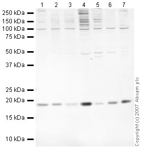
All lanes : Anti-JDP2 antibody (ab40916) at 1 µg/ml
Lane 1 : HeLa (Human epithelial carcinoma cell line) Whole Cell Lysate
Lane 2 : HeLa (Human epithelial carcinoma cell line) Nuclear Lysate
Lane 3 : Jurkat (Human T cell lymphoblast-like cell line) Whole Cell Lysate
Lane 4 : Jurkat nuclear extract lysate (ab14844)
Lane 5 :Hep G2 nuclear extract lysate (ab14660)
Lane 6 : Du145 (Human prostate carcinoma cell line) Whole Cell Lysate
Lane 7 : LNCaP (Human prostate adenocarcinoma cell line) Nuclear lysate
Lysates/proteins at 10 µg per lane.
Secondary
All lanes : IRDye 680 Conjugated Goat Anti-Rabbit IgG (H+L) at 1/10000 dilution
Performed under reducing conditions.
Predicted band size: 19 kDa
Observed band size: 19 kDa -
ICC/IF image of ab40916 stained HepG2 cells. The cells were 4% PFA fixed (10 min) and then incubated in 1%BSA / 10% normal goat serum / 0.3M glycine in 0.1% PBS-Tween for 1h to permeabilise the cells and block non-specific protein-protein interactions. The cells were then incubated with the antibody (ab40916, 1µg/ml) overnight at +4°C. The secondary antibody (green) was ab96899 Dylight® 488 goat anti-rabbit IgG (H+L) used at a 1/250 dilution for 1h. Alexa Fluor® 594 WGA was used to label plasma membranes (red) at a 1/200 dilution for 1h. DAPI was used to stain the cell nuclei (blue) at a concentration of 1.43µM.
-
IHC image of ab40916 staining in human cervix formalin fixed paraffin embedded tissue section, performed on a Leica BondTM system using the standard protocol F. The section was pre-treated using heat mediated antigen retrieval with sodium citrate buffer (pH6, epitope retrieval solution 1) for 20 mins. The section was then incubated with ab40916, 1µg/ml, for 15 mins at room temperature and detected using an HRP conjugated compact polymer system. DAB was used as the chromogen. The section was then counterstained with haematoxylin and mounted with DPX.
For other IHC staining systems (automated and non-automated) customers should optimize variable parameters such as antigen retrieval conditions, primary antibody concentration and antibody incubation times.
Protocols
References (5)
ab40916 has been referenced in 5 publications.
- Cao L et al. Genotoxic stress-triggered ß-catenin/JDP2/PRMT5 complex facilitates reestablishing glutathione homeostasis. Nat Commun 10:3761 (2019). PubMed: 31434880
- Kong X et al. Defining UHRF1 Domains that Support Maintenance of Human Colon Cancer DNA Methylation and Oncogenic Properties. Cancer Cell 35:633-648.e7 (2019). PubMed: 30956060
- Mansour MR et al. JDP2: An oncogenic bZIP transcription factor in T cell acute lymphoblastic leukemia. J Exp Med 215:1929-1945 (2018). PubMed: 29941549
- Tanigawa S et al. Jun dimerization protein 2 is a critical component of the Nrf2/MafK complex regulating the response to ROS homeostasis. Cell Death Dis 4:e921 (2013). WB ; Human . PubMed: 24232097
- Chiou SS et al. Control of Oxidative Stress and Generation of Induced Pluripotent Stem Cell-like Cells by Jun Dimerization Protein 2. Cancers (Basel) 5:959-84 (2013). PubMed: 24202329
Images
-
All lanes : Anti-JDP2 antibody (ab40916) at 1 µg/ml
Lane 1 : HeLa (Human epithelial carcinoma cell line) Whole Cell Lysate
Lane 2 : HeLa (Human epithelial carcinoma cell line) Nuclear Lysate
Lane 3 : Jurkat (Human T cell lymphoblast-like cell line) Whole Cell Lysate
Lane 4 : Jurkat nuclear extract lysate (ab14844)
Lane 5 :Hep G2 nuclear extract lysate (ab14660)
Lane 6 : Du145 (Human prostate carcinoma cell line) Whole Cell Lysate
Lane 7 : LNCaP (Human prostate adenocarcinoma cell line) Nuclear lysate
Lysates/proteins at 10 µg per lane.
Secondary
All lanes : IRDye 680 Conjugated Goat Anti-Rabbit IgG (H+L) at 1/10000 dilution
Performed under reducing conditions.
Predicted band size: 19 kDa
Observed band size: 19 kDa -
ICC/IF image of ab40916 stained HepG2 cells. The cells were 4% PFA fixed (10 min) and then incubated in 1%BSA / 10% normal goat serum / 0.3M glycine in 0.1% PBS-Tween for 1h to permeabilise the cells and block non-specific protein-protein interactions. The cells were then incubated with the antibody (ab40916, 1µg/ml) overnight at +4°C. The secondary antibody (green) was ab96899 Dylight® 488 goat anti-rabbit IgG (H+L) used at a 1/250 dilution for 1h. Alexa Fluor® 594 WGA was used to label plasma membranes (red) at a 1/200 dilution for 1h. DAPI was used to stain the cell nuclei (blue) at a concentration of 1.43µM.
-
IHC image of ab40916 staining in human cervix formalin fixed paraffin embedded tissue section, performed on a Leica BondTM system using the standard protocol F. The section was pre-treated using heat mediated antigen retrieval with sodium citrate buffer (pH6, epitope retrieval solution 1) for 20 mins. The section was then incubated with ab40916, 1µg/ml, for 15 mins at room temperature and detected using an HRP conjugated compact polymer system. DAB was used as the chromogen. The section was then counterstained with haematoxylin and mounted with DPX.
For other IHC staining systems (automated and non-automated) customers should optimize variable parameters such as antigen retrieval conditions, primary antibody concentration and antibody incubation times.


