Anti-Histone H2A antibody (ab18975)
")
Key features and details
- Rabbit polyclonal to Histone H2A
- Suitable for: WB, IHC-P
- Reacts with: Human
- Isotype: IgG
Overview
-
Product name
Anti-Histone H2A antibody
See all Histone H2A primary antibodies -
Description
Rabbit polyclonal to Histone H2A -
Host species
Rabbit -
Specificity
ab18975 does not cross-react with other histones. -
Tested Applications & Species
See all applications and species dataApplication Species IHC-P HumanWB Human
-
Immunogen
Synthetic peptide:
SGRGKQGGKARAKAKTRSSRAG
, corresponding to N terminal amino acids 2-23 of Human Histone H2A


Properties
-
Form
Liquid -
Storage instructions
Shipped at 4°C. Store at +4°C short term (1-2 weeks). Upon delivery aliquot. Store at -20°C or -80°C. Avoid freeze / thaw cycle. -
Storage buffer
Preservative: 0.03% Proclin 300
Constituents: PBS, 30% Glycerol (glycerin, glycerine), 0.5% BSA, 0.015% EDTA -
 Concentration information loading...
 -
Purity
Immunogen affinity purified -
Clonality
Polyclonal -
Isotype
IgG
Images
-
Western blot - Anti-Histone H2A antibody (ab18975)Anti-Histone H2A antibody (ab18975) at 1 µg/ml + HeLa cell lysate
Predicted band size: 16 kDa
Observed band size: ~14 kDa why is the actual band size different from the predicted?
-
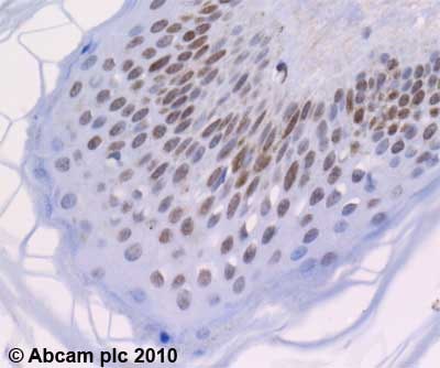 Immunohistochemistry (Formalin/PFA-fixed paraffin-embedded sections) - Anti-Histone H2A antibody (ab18975)ab18975 (2µg/ml) staining Histone H2A in human skin using an automated system (DAKO Autostainer Plus). Using this protocol there is nuclear staining in the epidermis.
Sections were rehydrated and antigen retrieved with the Dako 3 in 1 AR buffer citrate pH6.1 in a DAKO PT link. Slides were peroxidase blocked in 3% H2O2 in methanol for 10 mins. They were then blocked with Dako Protein block for 10 minutes (containing casein 0.25% in PBS) then incubated with primary antibody for 20 min and detected with Dako envision flex amplification kit for 30 minutes. Colorimetric detection was completed with Diaminobenzidine for 5 minutes. Slides were counterstained with Haematoxylin and coverslipped under DePeX. Please note that, for manual staining, optimization of primary antibody concentration and incubation time is recommended. Signal amplification may be required.


