Anti-HEXIM1 antibody (ab25388)
Key features and details
- Rabbit polyclonal to HEXIM1
- Suitable for: WB, ICC/IF, IP
- Reacts with: Human
- Isotype: IgG
Overview
-
Product name
Anti-HEXIM1 antibody
See all HEXIM1 primary antibodies -
Description
Rabbit polyclonal to HEXIM1 -
Host species
Rabbit -
Tested applications
Suitable for: WB, ICC/IF, IPmore details -
Species reactivity
Reacts with: Human
Predicted to work with: Mouse, Rat, Dog
-
Immunogen
Synthetic peptide conjugated to KLH derived from within residues 300 to the C-terminus of Human HEXIM1.
Read Abcam's proprietary immunogen policy (Peptide available as ab26978.)
Properties
-
Form
Liquid -
Storage instructions
Shipped at 4°C. Store at +4°C short term (1-2 weeks). Upon delivery aliquot. Store at -20°C or -80°C. Avoid freeze / thaw cycle. -
Storage buffer
pH: 7.40
Preservative: 0.02% Sodium azide
Constituent: PBS
Batches of this product that have a concentration Concentration information loading...
Purity
Immunogen affinity purifiedClonality
PolyclonalIsotype
IgGResearch areas
Associated products
-
ChIP Related Products
-
Compatible Secondaries
-
Isotype control
Applications
Our Abpromise guarantee covers the use of ab25388 in the following tested applications.
The application notes include recommended starting dilutions; optimal dilutions/concentrations should be determined by the end user.
Application Abreviews Notes WB Use a concentration of 1 µg/ml. Detects a band of approximately 54 kDa (predicted molecular weight: 41 kDa). Abcam recommends using milk (3%) as the blocking agent. ICC/IF Use a concentration of 1 µg/ml. IP Use at an assay dependent concentration. Target
-
Function
Transcriptional regulator which functions as a general RNA polymerase II transcription inhibitor. In cooperation with 7SK snRNA sequesters P-TEFb in a large inactive 7SK snRNP complex preventing RNA polymerase II phosphorylation and subsequent transcriptional elongation. May also regulate NF-kappa-B, ESR1, NR3C1 and CIITA-dependent transcriptional activity. -
Tissue specificity
Ubiquitously expressed with higher expression in placenta. HEXIM1 and HEXIM2 are differentially expressed. Expressed in endocrine tissues. -
Sequence similarities
Belongs to the HEXIM family. -
Domain
The coiled-coil domain mediates oligomerization. -
Cellular localization
Nucleus. Cytoplasm. Binds alpha-importin and is mostly nuclear. - Information by UniProt
-
Database links
- Entrez Gene: 10614 Human
- Entrez Gene: 192231 Mouse
- Entrez Gene: 498008 Rat
- Omim: 607328 Human
- SwissProt: O94992 Human
- SwissProt: Q8R409 Mouse
- SwissProt: Q5M9G1 Rat
- Unigene: 15299 Human
see all -
Alternative names
- Cardiac lineage protein 1 antibody
- CLP 1 antibody
- CLP1 antibody
see all
Images
-
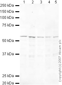
All lanes : Anti-HEXIM1 antibody (ab25388) at 1/250 dilution
Lane 1 : HeLa (Human epithelial carcinoma cell line) Whole Cell Lysate
Lane 2 : Jurkat (Human T cell lymphoblast-like cell line) Whole Cell Lysate
Lane 3 : A431 (Human epithelial carcinoma cell line) Whole Cell Lysate
Lane 4 : MCF7 (Human breast adenocarcinoma cell line) Whole Cell Lysate
Lane 5 : SHSY-5Y (Human neuroblastoma cell line) Whole Cell Lysate
Lysates/proteins at 10 µg per lane.
Secondary
All lanes : IRDye 680 Conjugated Goat Anti-Rabbit IgG (H+L) at 1/10000 dilution
Predicted band size: 41 kDa
Observed band size: 54 kDa why is the actual band size different from the predicted?
ab25388 recognizes a band at approximately 54 kDa that corresponds in size to that seen for HEXIM1. Although it has a predicted molecular weight of 41 kDa, it has been shown to migrate at a larger size of about 54-60 kDa (see Byers et al., J Biol Chem. 2005 Apr 22;280(16):16360-7 and Schulte et al., J. Biol. Chem., Vol. 280, (26): 24968-24977). -
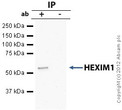
HEXIM1 - ChIP Grade was immunoprecipitated using 0.5mg Jurkat whole cell extract, 5µg of Rabbit polyclonal to HEXIM1 and 50µl of protein G magnetic beads (+). No antibody was added to the control (-).
The antibody was incubated under agitation with Protein G beads for 10min, Jurkat whole cell extract lysate diluted in RIPA buffer was added to each sample and incubated for a further 10min under agitation.
Proteins were eluted by addition of 40µl SDS loading buffer and incubated for 10min at 70oC; 10µl of each sample was separated on a SDS PAGE gel, transferred to a nitrocellulose membrane, blocked with 5% BSA and probed with ab25388.
Secondary: Mouse monoclonal [SB62a] Secondary Antibody to Rabbit IgG light chain (HRP) (ab99697).
Band: 54kD: HEXIM1 . -
ICC/IF image of ab25388 stained HeLa cells. The cells were 4% formaldehyde fixed (10 min) and then incubated in 1%BSA / 10% normal goat serum / 0.3M glycine in 0.1% PBS-Tween for 1h to permeabilise the cells and block non-specific protein-protein interactions. The cells were then incubated with the antibody ab25388 at 1µg/ml overnight at +4°C. The secondary antibody (green) was DyLight® 488 goat anti- rabbit (ab96899) IgG (H+L) used at a 1/1000 dilution for 1h. Alexa Fluor® 594 WGA was used to label plasma membranes (red) at a 1/200 dilution for 1h. DAPI was used to stain the cell nuclei (blue) at a concentration of 1.43µM.
Protocols
References (23)
ab25388 has been referenced in 23 publications.
- Edwards DS et al. BRD4 Prevents R-Loop Formation and Transcription-Replication Conflicts by Ensuring Efficient Transcription Elongation. Cell Rep 32:108166 (2020). PubMed: 32966794
- Sikorski K et al. A high-throughput pipeline for validation of antibodies. Nat Methods 15:909-912 (2018). PubMed: 30377371
- Bowry A et al. BET Inhibition Induces HEXIM1- and RAD51-Dependent Conflicts between Transcription and Replication. Cell Rep 25:2061-2069.e4 (2018). PubMed: 30463005
- Faust TB et al. The HIV-1 Tat protein recruits a ubiquitin ligase to reorganize the 7SK snRNP for transcriptional activation. Elife 7:N/A (2018). PubMed: 29845934
- Egloff S et al. The 7SK snRNP associates with the little elongation complex to promote snRNA gene expression. EMBO J 36:934-948 (2017). PubMed: 28254838
Images
-
All lanes : Anti-HEXIM1 antibody (ab25388) at 1/250 dilution
Lane 1 : HeLa (Human epithelial carcinoma cell line) Whole Cell Lysate
Lane 2 : Jurkat (Human T cell lymphoblast-like cell line) Whole Cell Lysate
Lane 3 : A431 (Human epithelial carcinoma cell line) Whole Cell Lysate
Lane 4 : MCF7 (Human breast adenocarcinoma cell line) Whole Cell Lysate
Lane 5 : SHSY-5Y (Human neuroblastoma cell line) Whole Cell Lysate
Lysates/proteins at 10 µg per lane.
Secondary
All lanes : IRDye 680 Conjugated Goat Anti-Rabbit IgG (H+L) at 1/10000 dilution
Predicted band size: 41 kDa
Observed band size: 54 kDa why is the actual band size different from the predicted?
ab25388 recognizes a band at approximately 54 kDa that corresponds in size to that seen for HEXIM1. Although it has a predicted molecular weight of 41 kDa, it has been shown to migrate at a larger size of about 54-60 kDa (see Byers et al., J Biol Chem. 2005 Apr 22;280(16):16360-7 and Schulte et al., J. Biol. Chem., Vol. 280, (26): 24968-24977). -
HEXIM1 - ChIP Grade was immunoprecipitated using 0.5mg Jurkat whole cell extract, 5µg of Rabbit polyclonal to HEXIM1 and 50µl of protein G magnetic beads (+). No antibody was added to the control (-).
The antibody was incubated under agitation with Protein G beads for 10min, Jurkat whole cell extract lysate diluted in RIPA buffer was added to each sample and incubated for a further 10min under agitation.
Proteins were eluted by addition of 40µl SDS loading buffer and incubated for 10min at 70oC; 10µl of each sample was separated on a SDS PAGE gel, transferred to a nitrocellulose membrane, blocked with 5% BSA and probed with ab25388.
Secondary: Mouse monoclonal [SB62a] Secondary Antibody to Rabbit IgG light chain (HRP) (ab99697).
Band: 54kD: HEXIM1 . -
ICC/IF image of ab25388 stained HeLa cells. The cells were 4% formaldehyde fixed (10 min) and then incubated in 1%BSA / 10% normal goat serum / 0.3M glycine in 0.1% PBS-Tween for 1h to permeabilise the cells and block non-specific protein-protein interactions. The cells were then incubated with the antibody ab25388 at 1µg/ml overnight at +4°C. The secondary antibody (green) was DyLight® 488 goat anti- rabbit (ab96899) IgG (H+L) used at a 1/1000 dilution for 1h. Alexa Fluor® 594 WGA was used to label plasma membranes (red) at a 1/200 dilution for 1h. DAPI was used to stain the cell nuclei (blue) at a concentration of 1.43µM.


