Anti-Glutaminase C antibody (ab262717)
Key features and details
- Rabbit polyclonal to Glutaminase C
- Suitable for: IHC-P, WB, ICC/IF, IP
- Reacts with: Human
- Isotype: IgG
Overview
-
Product name
Anti-Glutaminase C antibody
See all Glutaminase C primary antibodies -
Description
Rabbit polyclonal to Glutaminase C -
Host species
Rabbit -
Tested applications
Suitable for: IHC-P, WB, ICC/IF, IPmore details -
Species reactivity
Reacts with: Human -
Immunogen
Recombinant fragment within Human Glutaminase C (C terminal). The exact sequence is proprietary.
Database link: O94925 -
Positive control
- ICC/IF: A549 cells. IHC-P: Human breast carcinoma tissue. WB: GLS1(GAC isoform specific) shRNA-transfected MDA-MB-231 whole cell extract. IP: Glutaminase C protein from MDA-MB-231 whole cell extract.
Properties
-
Form
Liquid -
Storage instructions
Shipped at 4°C. Store at +4°C short term (1-2 weeks). Upon delivery aliquot. Store at -20°C long term. Avoid freeze / thaw cycle. -
Storage buffer
pH: 7.00
Preservative: 0.025% Proclin 300
Constituents: 79% PBS, 20% Glycerol (glycerin, glycerine) -
 Concentration information loading...
 -
Purity
Immunogen affinity purified -
Clonality
Polyclonal -
Isotype
IgG
Images
-
All lanes : Anti-Glutaminase C antibody (ab262717) at 1/5000 dilution
Lane 1 : Non-transfected MDA-MB-231 whole cell extract
Lane 2 : GLS1(GAC isoform specific) shRNA-transfected MDA-MB-231 whole cell extract
Lysates/proteins at 30 µg per lane.
Predicted band size: 73 kDaThis antibody specifically recognizes GAC but not the KGA isoform.
7.5% SDS-PAGE.
-
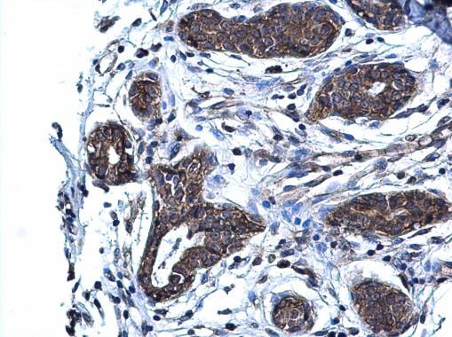
 Immunohistochemistry (Formalin/PFA-fixed paraffin-embedded sections) - Anti-Glutaminase C antibody (ab262717)
Paraffin embedded human breast carcinoma tissue stained for Glutaminase C using ab262717 at a 1/500 dilution in immunohistochemistry.
-
Immunoprecipitation of Glutaminase C protein from MDA-MB-231 whole cell extracts using 5 μg of ab262717.
Western blot analysis was performed using ab262717.
EasyBlot anti-Rabbit IgG was used as a secondary reagent.Lane 1: Input
Lane 2: Control IgG IP
Lane 3: ab262717 IP
-
A549 cells stained for Glutaminase C using ab262717 at a 1/500 dilution (green) in ICC/IF.
Cells were fixed in 4% paraformaldehyde at RT for 15 mins.
Red: Histone H3K9ac (acetyl Lys9), a nucleus marker, stained by Histone H3K9ac (acetyl Lys9) antibody diluted at 1/500.
Scale bar = 10 μm.


