Anti-Gli1 antibody (ab167388)
Key features and details
- Rabbit polyclonal to Gli1
- Suitable for: WB
- Reacts with: Mouse, Human
- Isotype: IgG
Overview
-
Product name
Anti-Gli1 antibody
See all Gli1 primary antibodies -
Description
Rabbit polyclonal to Gli1 -
Host species
Rabbit -
Tested Applications & Species
See all applications and species dataApplication Species WB MouseHuman -
Immunogen
-
Positive control
- This antibody gave a positive signal in the following Mouse tissue lysates: E14 Embryonic Brain; E16 Embryonic Brain; E18 Embryonic Brain.
Properties
-
Form
Liquid -
Storage instructions
Shipped at 4°C. Store at +4°C short term (1-2 weeks). Upon delivery aliquot. Store at -20°C or -80°C. Avoid freeze / thaw cycle. -
Storage buffer
pH: 7.40
Preservative: 0.02% Sodium azide
Constituent: PBS
Batches of this product that have a concentration Concentration information loading...
Purity
Immunogen affinity purifiedClonality
PolyclonalIsotype
IgGResearch areas
Associated products
-
Compatible Secondaries
-
Immunizing Peptide (Blocking)
-
Isotype control
-
Recombinant Protein
Applications
The Abpromise guarantee
Our Abpromise guarantee covers the use of ab167388 in the following tested applications.
The application notes include recommended starting dilutions; optimal dilutions/concentrations should be determined by the end user.
GuaranteedTested applications are guaranteed to work and covered by our Abpromise guarantee.
PredictedPredicted to work for this combination of applications and species but not guaranteed.
IncompatibleDoes not work for this combination of applications and species.
Application Species WB MouseHumanAll applications RatRabbitChimpanzeeMacaque monkeyGorillaChinese hamsterOrangutanApplication Abreviews Notes WB (1) Use a concentration of 1 µg/ml. Detects a band of approximately 118 kDa (predicted molecular weight: 118 kDa).Can be blocked with Mouse Gli1 peptide (ab201523).Notes WB
Use a concentration of 1 µg/ml. Detects a band of approximately 118 kDa (predicted molecular weight: 118 kDa).Can be blocked with Mouse Gli1 peptide (ab201523).Target
-
Function
Acts as a transcriptional activator. May regulate the transcription of specific genes during normal development. May play a role in craniofacial development and digital development, as well as development of the central nervous system and gastrointestinal tract. Mediates SHH signaling and thus cell proliferation and differentiation. -
Tissue specificity
Testis, myometrium and fallopian tube. Also expressed in the brain with highest expression in the cerebellum, optic nerve and olfactory tract. -
Sequence similarities
Belongs to the GLI C2H2-type zinc-finger protein family.
Contains 5 C2H2-type zinc fingers. -
Post-translational
modificationsPhosphorylated in vitro by ULK3. -
Cellular localization
Cytoplasm. Nucleus. Tethered in the cytoplasm by binding to SUFU. Activation and translocation to the nucleus is promoted by interaction with STK36. Phosphorylation by ULK3 may promote nuclear localization. Translocation to the nucleus is promoted by interaction with ZIC1. - Information by UniProt
-
Database links
- Entrez Gene: 2735 Human
- Entrez Gene: 14632 Mouse
- Entrez Gene: 140589 Rat
- Omim: 165220 Human
- SwissProt: P08151 Human
- SwissProt: P47806 Mouse
- Unigene: 632702 Human
- Unigene: 391450 Mouse
see all -
Alternative names
- Gli 1 antibody
- GLI antibody
- GLI family zinc finger 1 antibody
see all
Images
-
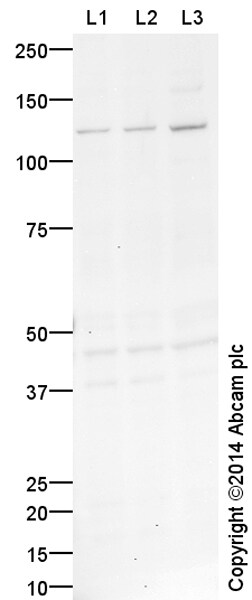
All lanes : Anti-Gli1 antibody (ab167388) at 1 µg/ml
Lane 1 : E14 Mouse Embryo Brain Tissue Lysate
Lane 2 : E16 Ms Embryo Brain Tissue Lysate
Lane 3 : E18 Ms Embryo Brain Tissue Lysate
Lysates/proteins at 20 µg per lane.
Secondary
All lanes : Goat Anti-Rabbit IgG H&L (HRP) (ab97051) at 1/50000 dilution
Developed using the ECL technique.
Performed under reducing conditions.
Predicted band size: 118 kDa
Observed band size: 118 kDa
Additional bands at: 45 kDa (possible non-specific binding)
Exposure time: 20 minutesThis blot was produced using a 4-12% Bis-tris gel under the MOPS buffer system. The gel was run at 200V for 50 minutes before being transferred onto a Nitrocellulose membrane at 30V for 70 minutes. The membrane was then blocked for an hour using 2% Bovine Serum Albumin before being incubated with ab167388 overnight at 4°C. Antibody binding was detected using an anti-rabbit antibody conjugated to HRP, and visualised using ECL development solution ab133406
Datasheets and documents
References (0)
ab167388 has not yet been referenced specifically in any publications.
Images
-
All lanes : Anti-Gli1 antibody (ab167388) at 1 µg/ml
Lane 1 : E14 Mouse Embryo Brain Tissue Lysate
Lane 2 : E16 Ms Embryo Brain Tissue Lysate
Lane 3 : E18 Ms Embryo Brain Tissue Lysate
Lysates/proteins at 20 µg per lane.
Secondary
All lanes : Goat Anti-Rabbit IgG H&L (HRP) (ab97051) at 1/50000 dilution
Developed using the ECL technique.
Performed under reducing conditions.
Predicted band size: 118 kDa
Observed band size: 118 kDa
Additional bands at: 45 kDa (possible non-specific binding)
Exposure time: 20 minutesThis blot was produced using a 4-12% Bis-tris gel under the MOPS buffer system. The gel was run at 200V for 50 minutes before being transferred onto a Nitrocellulose membrane at 30V for 70 minutes. The membrane was then blocked for an hour using 2% Bovine Serum Albumin before being incubated with ab167388 overnight at 4°C. Antibody binding was detected using an anti-rabbit antibody conjugated to HRP, and visualised using ECL development solution ab133406


