Anti-Giantin antibody - Golgi Marker (ab80864)
Key features and details
- Rabbit polyclonal to Giantin - Golgi Marker
- Suitable for: IHC-P, ICC/IF, WB
- Knockout validated
- Reacts with: Human
- Isotype: IgG
Overview
-
Product name
Anti-Giantin antibody - Golgi Marker
See all Giantin primary antibodies -
Description
Rabbit polyclonal to Giantin - Golgi Marker -
Host species
Rabbit -
Tested Applications & Species
See all applications and species dataApplication Species ICC/IF HumanIHC-P HumanWB Human -
Immunogen
Synthetic peptide corresponding to Human Giantin aa 1250-1350 conjugated to keyhole limpet haemocyanin.
(Peptide available asab101412) -
Positive control
- This antibody gave a positive signal in cervival tissue and MCF7 whole cells using immunohistochemistry and immunocytochemistry, respectively.
Properties
-
Form
Liquid -
Storage instructions
Shipped at 4°C. Store at +4°C short term (1-2 weeks). Upon delivery aliquot. Store at -20°C or -80°C. Avoid freeze / thaw cycle. -
Storage buffer
pH: 7.40
Preservative: 0.02% Sodium azide
Constituent: PBS
Batches of this product that have a concentration Concentration information loading...
Purity
Immunogen affinity purifiedClonality
PolyclonalIsotype
IgGResearch areas
Associated products
-
Compatible Secondaries
-
Isotype control
-
Recombinant Protein
-
Related Products
Applications
The Abpromise guarantee
Our Abpromise guarantee covers the use of ab80864 in the following tested applications.
The application notes include recommended starting dilutions; optimal dilutions/concentrations should be determined by the end user.
GuaranteedTested applications are guaranteed to work and covered by our Abpromise guarantee.
PredictedPredicted to work for this combination of applications and species but not guaranteed.
IncompatibleDoes not work for this combination of applications and species.
Application Species ICC/IF HumanIHC-P HumanWB HumanApplication Abreviews Notes IHC-P (1) Use a concentration of 0.1 - 1 µg/ml. Perform heat mediated antigen retrieval with citrate buffer pH 6 before commencing with IHC staining protocol.ICC/IF (1) Use a concentration of 1 µg/ml.WB 1/1000. Predicted molecular weight: 376 kDa.Notes IHC-P
Use a concentration of 0.1 - 1 µg/ml. Perform heat mediated antigen retrieval with citrate buffer pH 6 before commencing with IHC staining protocol.ICC/IF
Use a concentration of 1 µg/ml.WB
1/1000. Predicted molecular weight: 376 kDa.Target
-
Function
May participate in forming intercisternal cross-bridges of the Golgi complex. -
Cellular localization
Golgi apparatus membrane. - Information by UniProt
-
Database links
- Entrez Gene: 2804 Human
- Omim: 602500 Human
- SwissProt: Q14789 Human
- Unigene: 213389 Human
-
Alternative names
- 372 kDa Golgi complex associated protein antibody
- 372 kDa Golgi complex-associated protein antibody
- GCP antibody
see all
Images
-
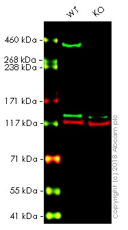
All lanes : Anti-Giantin antibody - Golgi Marker (ab80864) at 1/1000 dilution
Lane 1 : Wild-type HAP1 whole cell lysate
Lane 2 : GOLGB1 knockout HAP1 whole cell lysate
Lysates/proteins at 20 µg per lane.
Predicted band size: 376 kDaLanes 1 - 2: Merged signal (red and green). Green - ab80864 observed at 377kDa. Red - loading control, ab130007, observed at 130kDa.
ab80864 was shown to recognize GOLGB1 in wild-type HAP1 cells as signal was lost at the expected MW in GOLGB1 knockout cells. Additional cross-reactive bands were observed in the wild-type and knockout cells. Wild-type and GOLGB1 knockout samples were subjected to SDS-PAGE. Ab80864 and ab130007 (Mouse anti-Vinculin loading control) were incubated overnight at 4°C at 1/1000 dilution and 1/20000 dilution respectively. Blots were developed with Goat anti-Rabbit IgG H&L (IRDye® 800CW) preabsorbed ab216773 and Goat anti-Mouse IgG H&L (IRDye® 680RD) preabsorbed ab216776 secondary antibodies at 1/20000 dilution for 1 hour at room temperature before imaging.
-
ab80864 stained in Hela cells. Cells were fixed with 100% methanol (5min) at room temperature and incubated with PBS containing 10% goat serum, 0.3 M glycine, 1% BSA and 0.1% triton for 1h at room temperature to permeabilise the cells and block non-specific protein-protein interactions. The cells were then incubated with the antibody ab80864 at 1µg/ml and ab7291 (Mouse monoclonal [DM1A] to alpha Tubulin - Loading Control) at 1/1000 dilution overnight at +4°C. The secondary antibodies were ab150120 (pseudo-colored red) and ab150081 (colored green) used at 1 ug/ml for 1hour at room temperature. DAPI was used to stain the cell nuclei (colored blue) at a concentration of 1.43µM for 1hour at room temperature.
-
 Immunohistochemistry (Formalin/PFA-fixed paraffin-embedded sections) - Anti-Giantin antibody - Golgi Marker (ab80864)
IHC image of ab80864 staining in human cervix carcinoma formalin fixed paraffin embedded tissue section*, performed on a Leica BondTM system using the standard protocol F. The section was pre-treated using heat mediated antigen retrieval with sodium citrate buffer (pH6, epitope retrieval solution 1) for 20 mins. The section was then incubated with ab80864, 0.1µg/ml for 15 mins at room temperature and detected using an HRP conjugated compact polymer system. DAB was used as the chromogen. The section was then counterstained with haematoxylin and mounted with DPX
For other IHC staining systems (automated and non-automated) customers should optimize variable parameters such as antigen retrieval conditions, primary antibody concentration and antibody incubation times.
*Tissue obtained from the Human Research Tissue Bank, supported by the NIHR Cambridge Biomedical Research Centre
-
 Immunocytochemistry/ Immunofluorescence - Anti-Giantin antibody - Golgi Marker (ab80864)This image is courtesy of an anonymous Abreview
Ab80864 staining Giantin in WIF-B cells (hepatoma-derived hybrid cell line) by ICC/IF (Immunocytochemistry/Immunofluorescence). Cells were fixed with formaldehyde; permeabilized with 0.2% Triton X-100 and blocked with 1% Donkey serum in 0.1% PBST for 1 hour at 22°C. Samples were incubated at 1/25 dilution for 3 hours at 22°C. An Alexa Fluor® 594 Donkey anti rabbit was used as the secondary antibody at 1/200 dilution.
Protocols
References (14)
ab80864 has been referenced in 14 publications.
- Wang Y et al. Generation of a caged lentiviral vector through an unnatural amino acid for photo-switchable transduction. Nucleic Acids Res N/A:N/A (2019). PubMed: 31361892
- Yarur HE et al. Type 2ß Corticotrophin Releasing Factor Receptor Forms a Heteromeric Complex With Dopamine D1 Receptor in Living Cells. Front Pharmacol 10:1501 (2019). PubMed: 31969820
- Ishida M & Bonifacino JS ARFRP1 functions upstream of ARL1 and ARL5 to coordinate recruitment of distinct tethering factors to the trans-Golgi network. J Cell Biol 218:3681-3696 (2019). PubMed: 31575603
- Bachelot A et al. A common African variant of human connexin 37 is associated with Caucasian primary ovarian insufficiency and has a deleterious effect in vitro. Int J Mol Med 41:640-648 (2018). PubMed: 29207017
- Tian S et al. Genome-wide CRISPR screens for Shiga toxins and ricin reveal Golgi proteins critical for glycosylation. PLoS Biol 16:e2006951 (2018). PubMed: 30481169
Images
-
All lanes : Anti-Giantin antibody - Golgi Marker (ab80864) at 1/1000 dilution
Lane 1 : Wild-type HAP1 whole cell lysate
Lane 2 : GOLGB1 knockout HAP1 whole cell lysate
Lysates/proteins at 20 µg per lane.
Predicted band size: 376 kDaLanes 1 - 2: Merged signal (red and green). Green - ab80864 observed at 377kDa. Red - loading control, ab130007, observed at 130kDa.
ab80864 was shown to recognize GOLGB1 in wild-type HAP1 cells as signal was lost at the expected MW in GOLGB1 knockout cells. Additional cross-reactive bands were observed in the wild-type and knockout cells. Wild-type and GOLGB1 knockout samples were subjected to SDS-PAGE. Ab80864 and ab130007 (Mouse anti-Vinculin loading control) were incubated overnight at 4°C at 1/1000 dilution and 1/20000 dilution respectively. Blots were developed with Goat anti-Rabbit IgG H&L (IRDye® 800CW) preabsorbed ab216773 and Goat anti-Mouse IgG H&L (IRDye® 680RD) preabsorbed ab216776 secondary antibodies at 1/20000 dilution for 1 hour at room temperature before imaging.
-
ab80864 stained in Hela cells. Cells were fixed with 100% methanol (5min) at room temperature and incubated with PBS containing 10% goat serum, 0.3 M glycine, 1% BSA and 0.1% triton for 1h at room temperature to permeabilise the cells and block non-specific protein-protein interactions. The cells were then incubated with the antibody ab80864 at 1µg/ml and ab7291 (Mouse monoclonal [DM1A] to alpha Tubulin - Loading Control) at 1/1000 dilution overnight at +4°C. The secondary antibodies were ab150120 (pseudo-colored red) and ab150081 (colored green) used at 1 ug/ml for 1hour at room temperature. DAPI was used to stain the cell nuclei (colored blue) at a concentration of 1.43µM for 1hour at room temperature.
-
 Immunohistochemistry (Formalin/PFA-fixed paraffin-embedded sections) - Anti-Giantin antibody - Golgi Marker (ab80864)
IHC image of ab80864 staining in human cervix carcinoma formalin fixed paraffin embedded tissue section*, performed on a Leica BondTM system using the standard protocol F. The section was pre-treated using heat mediated antigen retrieval with sodium citrate buffer (pH6, epitope retrieval solution 1) for 20 mins. The section was then incubated with ab80864, 0.1µg/ml for 15 mins at room temperature and detected using an HRP conjugated compact polymer system. DAB was used as the chromogen. The section was then counterstained with haematoxylin and mounted with DPX
For other IHC staining systems (automated and non-automated) customers should optimize variable parameters such as antigen retrieval conditions, primary antibody concentration and antibody incubation times.
*Tissue obtained from the Human Research Tissue Bank, supported by the NIHR Cambridge Biomedical Research Centre
-
 Immunocytochemistry/ Immunofluorescence - Anti-Giantin antibody - Golgi Marker (ab80864) This image is courtesy of an anonymous Abreview
Ab80864 staining Giantin in WIF-B cells (hepatoma-derived hybrid cell line) by ICC/IF (Immunocytochemistry/Immunofluorescence). Cells were fixed with formaldehyde; permeabilized with 0.2% Triton X-100 and blocked with 1% Donkey serum in 0.1% PBST for 1 hour at 22°C. Samples were incubated at 1/25 dilution for 3 hours at 22°C. An Alexa Fluor® 594 Donkey anti rabbit was used as the secondary antibody at 1/200 dilution.


