Anti-EGFR (phospho Y845) antibody (ab109685)
 antibody (ab109685)")
Key features and details
- Rabbit polyclonal to EGFR (phospho Y845)
- Suitable for: ICC/IF, WB
- Reacts with: Human
- Isotype: IgG
Overview
-
Product name
Anti-EGFR (phospho Y845) antibody
See all EGFR primary antibodies -
Description
Rabbit polyclonal to EGFR (phospho Y845) -
Host species
Rabbit -
Tested applications
Suitable for: ICC/IF, WBmore details -
Species reactivity
Reacts with: Human
Predicted to work with: Mouse, Rat, Rabbit, Horse, Pig, Chimpanzee, Macaque monkey, Orangutan
-
Immunogen
Synthetic peptide. This information is proprietary to Abcam and/or its suppliers.
-
Positive control
- This antibody gave a positive signal in both EGF treated A431 and W138 whole cell lysates. This antibody gave a positive result in IF in the following cell lines:HeLa.
Properties
-
Form
Liquid -
Storage instructions
Shipped at 4°C. Store at +4°C short term (1-2 weeks). Upon delivery aliquot. Store at -20°C or -80°C. Avoid freeze / thaw cycle. -
Storage buffer
pH: 7.40
Preservative: 0.02% Sodium azide
Constituent: PBS
Batches of this product that have a concentration Concentration information loading...
Purity
Immunogen affinity purifiedClonality
PolyclonalIsotype
IgGResearch areas
Associated products
-
Compatible Secondaries
-
Isotype control
Applications
Our Abpromise guarantee covers the use of ab109685 in the following tested applications.
The application notes include recommended starting dilutions; optimal dilutions/concentrations should be determined by the end user.
Application Abreviews Notes ICC/IF Use a concentration of 10 µg/ml. WB Use a concentration of 1 µg/ml. Detects a band of approximately 190 kDa (predicted molecular weight: 134 kDa). Target
-
Function
Receptor tyrosine kinase binding ligands of the EGF family and activating several signaling cascades to convert extracellular cues into appropriate cellular responses. Known ligands include EGF, TGFA/TGF-alpha, amphiregulin, epigen/EPGN, BTC/betacellulin, epiregulin/EREG and HBEGF/heparin-binding EGF. Ligand binding triggers receptor homo- and/or heterodimerization and autophosphorylation on key cytoplasmic residues. The phosphorylated receptor recruits adapter proteins like GRB2 which in turn activates complex downstream signaling cascades. Activates at least 4 major downstream signaling cascades including the RAS-RAF-MEK-ERK, PI3 kinase-AKT, PLCgamma-PKC and STATs modules. May also activate the NF-kappa-B signaling cascade. Also directly phosphorylates other proteins like RGS16, activating its GTPase activity and probably coupling the EGF receptor signaling to the G protein-coupled receptor signaling. Also phosphorylates MUC1 and increases its interaction with SRC and CTNNB1/beta-catenin.
Isoform 2 may act as an antagonist of EGF action. -
Tissue specificity
Ubiquitously expressed. Isoform 2 is also expressed in ovarian cancers. -
Involvement in disease
Lung cancer
Inflammatory skin and bowel disease, neonatal, 2 -
Sequence similarities
Belongs to the protein kinase superfamily. Tyr protein kinase family. EGF receptor subfamily.
Contains 1 protein kinase domain. -
Post-translational
modificationsPhosphorylation at Ser-695 is partial and occurs only if Thr-693 is phosphorylated. Phosphorylation at Thr-678 and Thr-693 by PRKD1 inhibits EGF-induced MAPK8/JNK1 activation. Dephosphorylation by PTPRJ prevents endocytosis and stabilizes the receptor at the plasma membrane. Autophosphorylation at Tyr-1197 is stimulated by methylation at Arg-1199 and enhances interaction with PTPN6. Autophosphorylation at Tyr-1092 and/or Tyr-1110 recruits STAT3. Dephosphorylated by PTPN1 and PTPN2.
Monoubiquitinated and polyubiquitinated upon EGF stimulation; which does not affect tyrosine kinase activity or signaling capacity but may play a role in lysosomal targeting. Polyubiquitin linkage is mainly through 'Lys-63', but linkage through 'Lys-48', 'Lys-11' and 'Lys-29' also occurs. Deubiquitination by OTUD7B prevents degradation. Ubiquitinated by RNF115 and RNF126.
Methylated. Methylation at Arg-1199 by PRMT5 stimulates phosphorylation at Tyr-1197. -
Cellular localization
Secreted and Cell membrane. Endoplasmic reticulum membrane. Golgi apparatus membrane. Nucleus membrane. Endosome. Endosome membrane. Nucleus. In response to EGF, translocated from the cell membrane to the nucleus via Golgi and ER. Endocytosed upon activation by ligand. Colocalized with GPER1 in the nucleus of estrogen agonist-induced cancer-associated fibroblasts (CAF). - Information by UniProt
-
Database links
- Entrez Gene: 1956 Human
- Entrez Gene: 13649 Mouse
- Entrez Gene: 397070 Pig
- Entrez Gene: 24329 Rat
- Omim: 131550 Human
- SwissProt: P00533 Human
- SwissProt: Q01279 Mouse
- Unigene: 488293 Human
see all -
Alternative names
- Avian erythroblastic leukemia viral (v erb b) oncogene homolog antibody
- Cell growth inhibiting protein 40 antibody
- Cell proliferation inducing protein 61 antibody
see all
Images
-
Western blot - Anti-EGFR (phospho Y845) antibody (ab109685)All lanes : Anti-EGFR (phospho Y845) antibody (ab109685) at 1 µg/ml
Lane 1 : EGF-Stimulated A431 Whole Cell Lysate
Lane 2 :WI-38 whole cell lysate (ab3960)
Lane 3 : EGF-Stimulated A431 Whole Cell Lysate with Immunizing peptide at 1 µg/ml
Lane 4 :WI-38 whole cell lysate (ab3960) with Immunizing peptide at 1 µg/ml
Lysates/proteins at 10 µg per lane.
Secondary
All lanes : Goat Anti-Rabbit IgG H&L (HRP) preadsorbed (ab97080) at 1/5000 dilution
Developed using the ECL technique.
Performed under reducing conditions.
Predicted band size: 134 kDa
Observed band size: 190 kDa why is the actual band size different from the predicted?
Additional bands at: 125 kDa. We are unsure as to the identity of these extra bands.
Exposure time: 12 minutes
EGFR contains a number of potential glycosylation sites (SwissProt) which may explain its migration at a higher molecular weight than predicted. -
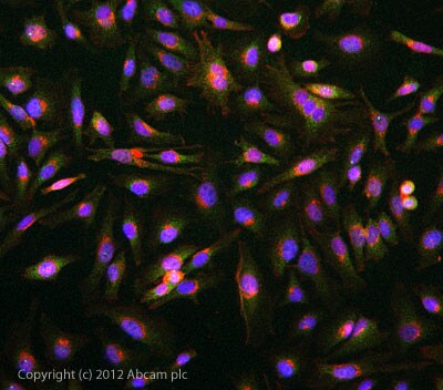 Immunocytochemistry/ Immunofluorescence - Anti-EGFR (phospho Y845) antibody (ab109685)
ICC/IF image of ab109685 stained HeLa cells. The cells were 4% formaldehyde fixed (10 min) and then incubated in 1%BSA / 10% normal goat serum / 0.3M glycine in 0.1% PBS-Tween for 1h to permeabilise the cells and block non-specific protein-protein interactions. The cells were then incubated with the antibody ab109685 at 10µg/ml overnight at +4°C. The secondary antibody (green) was DyLight® 488 goat anti- rabbit (ab96899) IgG (H+L) used at a 1/250 dilution for 1h. Alexa Fluor® 594 WGA was used to label plasma membranes (red) at a 1/200 dilution for 1h. DAPI was used to stain the cell nuclei (blue) at a concentration of 1.43µM.
Protocols
Datasheets and documents
References (0)
ab109685 has not yet been referenced specifically in any publications.
Images
-
Western blot - Anti-EGFR (phospho Y845) antibody (ab109685)All lanes : Anti-EGFR (phospho Y845) antibody (ab109685) at 1 µg/ml
Lane 1 : EGF-Stimulated A431 Whole Cell Lysate
Lane 2 :WI-38 whole cell lysate (ab3960)
Lane 3 : EGF-Stimulated A431 Whole Cell Lysate with Immunizing peptide at 1 µg/ml
Lane 4 :WI-38 whole cell lysate (ab3960) with Immunizing peptide at 1 µg/ml
Lysates/proteins at 10 µg per lane.
Secondary
All lanes : Goat Anti-Rabbit IgG H&L (HRP) preadsorbed (ab97080) at 1/5000 dilution
Developed using the ECL technique.
Performed under reducing conditions.
Predicted band size: 134 kDa
Observed band size: 190 kDa why is the actual band size different from the predicted?
Additional bands at: 125 kDa. We are unsure as to the identity of these extra bands.
Exposure time: 12 minutes
EGFR contains a number of potential glycosylation sites (SwissProt) which may explain its migration at a higher molecular weight than predicted. -
Immunocytochemistry/ Immunofluorescence - Anti-EGFR (phospho Y845) antibody (ab109685)
ICC/IF image of ab109685 stained HeLa cells. The cells were 4% formaldehyde fixed (10 min) and then incubated in 1%BSA / 10% normal goat serum / 0.3M glycine in 0.1% PBS-Tween for 1h to permeabilise the cells and block non-specific protein-protein interactions. The cells were then incubated with the antibody ab109685 at 10µg/ml overnight at +4°C. The secondary antibody (green) was DyLight® 488 goat anti- rabbit (ab96899) IgG (H+L) used at a 1/250 dilution for 1h. Alexa Fluor® 594 WGA was used to label plasma membranes (red) at a 1/200 dilution for 1h. DAPI was used to stain the cell nuclei (blue) at a concentration of 1.43µM.


