Anti-CFTR antibody (ab131553)
Key features and details
- Rabbit polyclonal to CFTR
- Suitable for: IHC-P, ICC
- Reacts with: Human
- Isotype: IgG
Overview
-
Product name
Anti-CFTR antibody
See all CFTR primary antibodies -
Description
Rabbit polyclonal to CFTR -
Host species
Rabbit -
Tested applications
Suitable for: IHC-P, ICCmore details -
Species reactivity
Reacts with: Human
Predicted to work with: Sheep, Rabbit, Cow, Pig, Chimpanzee, Macaque monkey, Gorilla
-
Immunogen
Synthetic peptide corresponding to Human CFTR aa 1250-1350 conjugated to keyhole limpet haemocyanin.
(Peptide available asab166746) -
Positive control
- This antibody gave a positive signal in Human normal lung formalin fixed paraffin embedded tissue section within IHC-P as well as in HCT116 and within ICC/IF.
-
General notes
Reproducibility is key to advancing scientific discovery and accelerating scientists’ next breakthrough.
Abcam is leading the way with our range of recombinant antibodies, knockout-validated antibodies and knockout cell lines, all of which support improved reproducibility.
We are also planning to innovate the way in which we present recommended applications and species on our product datasheets, so that only applications & species that have been tested in our own labs, our suppliers or by selected trusted collaborators are covered by our Abpromise™ guarantee.
In preparation for this, we have started to update the applications & species that this product is Abpromise guaranteed for.
We are also updating the applications & species that this product has been “predicted to work with,” however this information is not covered by our Abpromise guarantee.
Applications & species from publications and Abreviews that have not been tested in our own labs or in those of our suppliers are not covered by the Abpromise guarantee.
Please check that this product meets your needs before purchasing. If you have any questions, special requirements or concerns, please send us an inquiry and/or contact our Support team ahead of purchase. Recommended alternatives for this product can be found below, as well as customer reviews and Q&As.
Properties
-
Form
Liquid -
Storage instructions
Shipped at 4°C. Store at +4°C short term (1-2 weeks). Upon delivery aliquot. Store at -20°C or -80°C. Avoid freeze / thaw cycle. -
Storage buffer
pH: 7.40
Preservative: 0.02% Sodium azide
Constituent: PBS
Batches of this product that have a concentration Concentration information loading...
Purity
Immunogen affinity purifiedClonality
PolyclonalIsotype
IgGResearch areas
Associated products
-
Compatible Secondaries
-
Isotype control
-
Recombinant Protein
Applications
Our Abpromise guarantee covers the use of ab131553 in the following tested applications.
The application notes include recommended starting dilutions; optimal dilutions/concentrations should be determined by the end user.
Application Abreviews Notes IHC-P Use a concentration of 1 µg/ml. Perform heat mediated antigen retrieval with citrate buffer pH 6 before commencing with IHC staining protocol. ICC Use a concentration of 5 µg/ml. Target
-
Function
Involved in the transport of chloride ions. May regulate bicarbonate secretion and salvage in epithelial cells by regulating the SLC4A7 transporter. -
Tissue specificity
Found on the surface of the epithelial cells that line the lungs and other organs. -
Involvement in disease
Defects in CFTR are the cause of cystic fibrosis (CF) [MIM:219700]; also known as mucoviscidosis. CF is the most common genetic disease in the Caucasian population, with a prevalence of about 1 in 2'000 live births. Inheritance is autosomal recessive. CF is a common generalized disorder of exocrine gland function which impairs clearance of secretions in a variety of organs. It is characterized by the triad of chronic bronchopulmonary disease (with recurrent respiratory infections), pancreatic insufficiency (which leads to malabsorption and growth retardation) and elevated sweat electrolytes.
Defects in CFTR are the cause of congenital bilateral absence of the vas deferens (CBAVD) [MIM:277180]. CBAVD is an important cause of sterility in men and could represent an incomplete form of cystic fibrosis, as the majority of men suffering from cystic fibrosis lack the vas deferens. -
Sequence similarities
Belongs to the ABC transporter superfamily. ABCC family. CFTR transporter (TC 3.A.1.202) subfamily.
Contains 2 ABC transmembrane type-1 domains.
Contains 2 ABC transporter domains. -
Domain
The PDZ-binding motif mediates interactions with GOPC and with the SLC4A7, SLC9A3R1/EBP50 complex. -
Post-translational
modificationsPhosphorylated; activates the channel. It is not clear whether PKC phosphorylation itself activates the channel or permits activation by phosphorylation at PKA sites.
Ubiquitinated, leading to its degradation in the lysosome. Deubiquitination by USP10 in early endosomes, enhances its endocytic recycling. -
Cellular localization
Early endosome membrane. - Information by UniProt
-
Database links
- Entrez Gene: 463674 Chimpanzee
- Entrez Gene: 281067 Cow
- Entrez Gene: 101132066 Gorilla
- Entrez Gene: 1080 Human
- Entrez Gene: 403154 Pig
- Entrez Gene: 100009471 Rabbit
- Entrez Gene: 443347 Sheep
- Omim: 602421 Human
see all -
Alternative names
- ABC 35 antibody
- ABC35 antibody
- ABCC 7 antibody
see all
Images
-
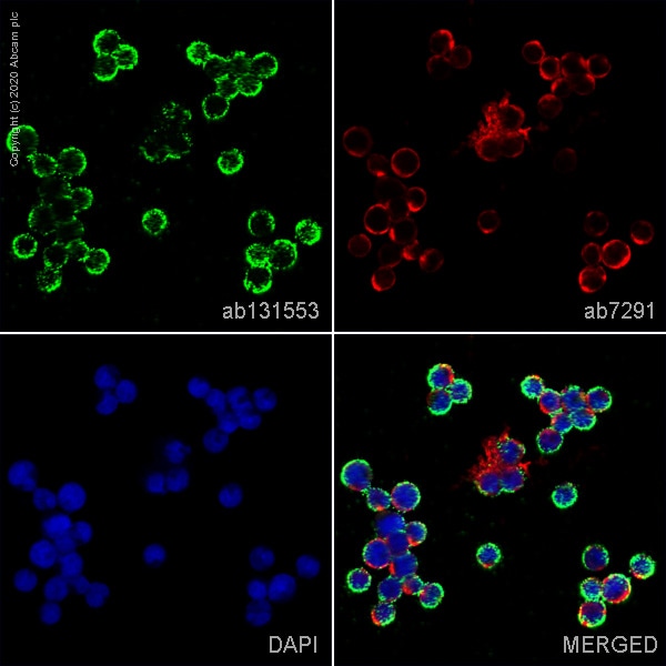
ab131553 staining Cystic fibrosis transmembrane conductance regulator in MOLT4 cells. The cells were fixed with 100% methanol (5 min), permeabilized with 0.1% PBS-Tween for 5 minutes and then blocked with 1% BSA/10% normal goat serum/0.3M glycine in 0.1%PBS-Tween for 1h. The cells were then incubated overnight at 4°C with ab131553 at 5 µg/ml and ab7291, Mouse monoclonal [DM1A] to alpha Tubulin - Loading Control. Cells were then incubated with ab150081, Goat polyclonal Secondary Antibody to Rabbit IgG - H&L (Alexa Fluor® 488), pre-adsorbed at 1/1000 dilution (shown in green) and ab150120, Goat polyclonal Secondary Antibody to Mouse IgG - H&L (Alexa Fluor® 594), pre-adsorbed at 1/1000 dilution (shown in pseudocolour red). Nuclear DNA was labelled with DAPI (shown in blue).
Image was acquired with a confocal microscope (Leica-Microsystems TCS SP8) and a single confocal section is shown.
-
 Immunohistochemistry (Formalin/PFA-fixed paraffin-embedded sections) - Anti-CFTR antibody (ab131553)
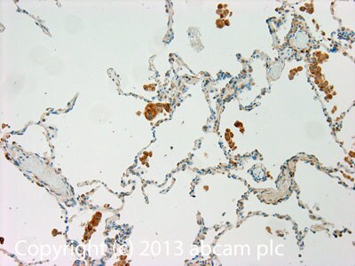
IHC image of CFTR staining in Human normal lung formalin fixed paraffin embedded tissue section, performed on a Leica BondTM system using the standard protocol F. The section was pre-treated using heat mediated antigen retrieval with sodium citrate buffer (pH6, epitope retrieval solution 1) for 20 mins. The section was then incubated with ab131553, 1µg/ml, for 15 mins at room temperature and detected using an HRP conjugated compact polymer system. DAB was used as the chromogen. The section was then counterstained with haematoxylin and mounted with DPX.
For other IHC staining systems (automated and non-automated) customers should optimize variable parameters such as antigen retrieval conditions, primary antibody concentration and antibody incubation times. -
ICC/IF image of ab131553 stained HCT116 cells. The cells were 100% methanol fixed (5 min) and then incubated in 1%BSA / 10% normal goat serum / 0.3M glycine in 0.1% PBS-Tween for 1h to permeabilise the cells and block non-specific protein-protein interactions. The cells were then incubated with the antibody (ab96899, 5µg/ml) overnight at +4°C. The secondary antibody (green) was ab96899, DyLight® 488 goat anti-rabbit IgG (H+L) used at a 1/250 dilution for 1h. Alexa Fluor® 594 WGA was used to label plasma membranes (red) at a 1/200 dilution for 1h. DAPI was used to stain the cell nuclei (blue) at a concentration of 1.43µM.
Protocols
Datasheets and documents
References (5)
ab131553 has been referenced in 5 publications.
- Hennig A et al. CFTR Expression Analysis for Subtyping of Human Pancreatic Cancer Organoids. Stem Cells Int 2019:1024614 (2019). PubMed: 31191661
- Dunn VK & Gleason E Inhibition of endocytosis suppresses the nitric oxide-dependent release of Cl- in retinal amacrine cells. PLoS One 13:e0201184 (2018). PubMed: 30044876
- Krishnan V et al. A role for the cystic fibrosis transmembrane conductance regulator in the nitric oxide-dependent release of Cl- from acidic organelles in amacrine cells. J Neurophysiol 118:2842-2852 (2017). PubMed: 28835528
- Uchida H et al. A xenogeneic-free system generating functional human gut organoids from pluripotent stem cells. JCI Insight 2:e86492 (2017). PubMed: 28097227
- Philippe R et al. SERCA and PMCA pumps contribute to the deregulation of Ca2+ homeostasis in human CF epithelial cells. Biochim Biophys Acta 1853:892-903 (2015). PubMed: 25661196
Images
-
ab131553 staining Cystic fibrosis transmembrane conductance regulator in MOLT4 cells. The cells were fixed with 100% methanol (5 min), permeabilized with 0.1% PBS-Tween for 5 minutes and then blocked with 1% BSA/10% normal goat serum/0.3M glycine in 0.1%PBS-Tween for 1h. The cells were then incubated overnight at 4°C with ab131553 at 5 µg/ml and ab7291, Mouse monoclonal [DM1A] to alpha Tubulin - Loading Control. Cells were then incubated with ab150081, Goat polyclonal Secondary Antibody to Rabbit IgG - H&L (Alexa Fluor® 488), pre-adsorbed at 1/1000 dilution (shown in green) and ab150120, Goat polyclonal Secondary Antibody to Mouse IgG - H&L (Alexa Fluor® 594), pre-adsorbed at 1/1000 dilution (shown in pseudocolour red). Nuclear DNA was labelled with DAPI (shown in blue).
Image was acquired with a confocal microscope (Leica-Microsystems TCS SP8) and a single confocal section is shown.
-
 Immunohistochemistry (Formalin/PFA-fixed paraffin-embedded sections) - Anti-CFTR antibody (ab131553)
IHC image of CFTR staining in Human normal lung formalin fixed paraffin embedded tissue section, performed on a Leica BondTM system using the standard protocol F. The section was pre-treated using heat mediated antigen retrieval with sodium citrate buffer (pH6, epitope retrieval solution 1) for 20 mins. The section was then incubated with ab131553, 1µg/ml, for 15 mins at room temperature and detected using an HRP conjugated compact polymer system. DAB was used as the chromogen. The section was then counterstained with haematoxylin and mounted with DPX.
For other IHC staining systems (automated and non-automated) customers should optimize variable parameters such as antigen retrieval conditions, primary antibody concentration and antibody incubation times. -
ICC/IF image of ab131553 stained HCT116 cells. The cells were 100% methanol fixed (5 min) and then incubated in 1%BSA / 10% normal goat serum / 0.3M glycine in 0.1% PBS-Tween for 1h to permeabilise the cells and block non-specific protein-protein interactions. The cells were then incubated with the antibody (ab96899, 5µg/ml) overnight at +4°C. The secondary antibody (green) was ab96899, DyLight® 488 goat anti-rabbit IgG (H+L) used at a 1/250 dilution for 1h. Alexa Fluor® 594 WGA was used to label plasma membranes (red) at a 1/200 dilution for 1h. DAPI was used to stain the cell nuclei (blue) at a concentration of 1.43µM.


