Anti-Cdk9 antibody (ab38840)
Key features and details
- Rabbit polyclonal to Cdk9
- Suitable for: ICC/IF, Sandwich ELISA, IHC-P, WB
- Reacts with: Human
- Isotype: IgG
Overview
-
Product name
Anti-Cdk9 antibody
See all Cdk9 primary antibodies -
Description
Rabbit polyclonal to Cdk9 -
Host species
Rabbit -
Tested applications
Suitable for: ICC/IF, Sandwich ELISA, IHC-P, WBmore details -
Species reactivity
Reacts with: Human
Predicted to work with: Mouse, Rat, Cow
-
Immunogen
Synthetic peptide conjugated to KLH derived from within residues 350 to the C-terminus of Human Cdk9.
Read Abcam's proprietary immunogen policy (Peptide available as ab39096.) -
Positive control
- This antibody gave a positive signal in the following whole cell lysates: HeLa (Human epithelial carcinoma cell line) Jurkat (Human T cell lymphoblast-like cell line) A431 (Human epithelial carcinoma cell line) HEK 293 (Human embryonic kidney cell line)
Properties
-
Form
Liquid -
Storage instructions
Shipped at 4°C. Store at +4°C short term (1-2 weeks). Upon delivery aliquot. Store at -20°C or -80°C. Avoid freeze / thaw cycle. -
Storage buffer
pH: 7.40
Preservative: 0.02% Sodium azide
Constituent: PBS
Batches of this product that have a concentration Concentration information loading...
Purity
Immunogen affinity purifiedClonality
PolyclonalIsotype
IgGResearch areas
Associated products
-
Compatible Secondaries
-
Isotype control
-
Recombinant Protein
-
Related Products
Applications
Our Abpromise guarantee covers the use of ab38840 in the following tested applications.
The application notes include recommended starting dilutions; optimal dilutions/concentrations should be determined by the end user.
Application Abreviews Notes ICC/IF Use a concentration of 1 µg/ml. Sandwich ELISA Use a concentration of 0.5 µg/ml. For sandwich ELISA, use this antibody as Detection at 0.5 µg/ml with Mouse monoclonal to Cdk9 (ab76873) as Capture. IHC-P Use a concentration of 1 µg/ml. WB 1/250. Detects a band of approximately 41 kDa (predicted molecular weight: 43, 50 kDa). Target
-
Function
Member of the cyclin-dependent kinase pair (CDK9/cyclin-T) complex, also called positive transcription elongation factor b (P-TEFb), which facilitates the transition from abortive to production elongation by phosphorylating the CTD (C-terminal domain) of the large subunit of RNA polymerase II (RNAP II), SUPT5H and RDBP. The CDK9/cyclin-K complex has also a kinase activity toward CTD of RNAP II and can substitute for P-TEFb in vitro. -
Tissue specificity
Ubiquitous. -
Sequence similarities
Belongs to the protein kinase superfamily. CMGC Ser/Thr protein kinase family. CDC2/CDKX subfamily.
Contains 1 protein kinase domain. -
Cellular localization
Nucleus. - Information by UniProt
-
Database links
- Entrez Gene: 520580 Cow
- Entrez Gene: 1025 Human
- Entrez Gene: 107951 Mouse
- Entrez Gene: 362110 Rat
- Omim: 603251 Human
- SwissProt: Q5EAB2 Cow
- SwissProt: P50750 Human
- SwissProt: Q99J95 Mouse
see all -
Alternative names
- C-2K antibody
- CDC2 related kinase antibody
- CDC2L4 antibody
see all
Images
-
ICC/IF image of ab38840 stained human HeLa cells. The cells were PFA fixed (10 min), permabilised in TBS-T (20 min) and incubated with the antibody (ab38840, 1µg/ml) for 1h at room temperature. 1%BSA / 10% normal serum / 0.3M glycine was used to quench autofluorescence and block non-specific protein-protein interactions. The secondary antibody (green) was Alexa Fluor® 488 goat anti-rabbit IgG (H+L) used at a 1/1000 dilution for 1h. Alexa Fluor® 594 WGA was used to label plasma membranes (red). DAPI was used to stain the cell nuclei (blue).
-
Standard Curve for Cdk9 (Analyte: Cdk9 protein (Tagged) (ab85603)); dilution range 1pg/ml to 1µg/ml using Capture Antibody Mouse monoclonal to Cdk9 (ab76873) at 5µg/ml and Detector Antibody Rabbit polyclonal to Cdk9 (ab38840) at 0.5µg/ml.
-
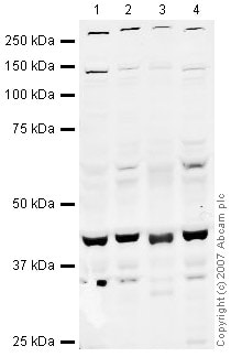
All lanes : Anti-Cdk9 antibody (ab38840) at 1/250 dilution
Lane 1 : HeLa (Human epithelial carcinoma cell line) Whole Cell Lysate
Lane 2 :Jurkat whole cell lysate (ab7899)
Lane 3 :A-431 whole cell lysate (ab7909)
Lane 4 :HEK-293 whole cell lysate (ab7902)
Lysates/proteins at 10 µg per lane.
Secondary
All lanes : IRDye 680 Conjugated Goat Anti-Rabbit IgG (H+L) at 1/10000 dilution
Performed under reducing conditions.
Predicted band size: 43, 50 kDa
Observed band size: 41 kDa why is the actual band size different from the predicted?
Additional bands at: 140 kDa, 260 kDa. We are unsure as to the identity of these extra bands.An additional band at approximately 34 kDa is occasionally observed with ab38840. This is thought to be due to cross-reactivity.
-
IHC image of ab38840 staining Cdk9 in Human breast adenocarcinoma formalin fixed paraffin embedded tissue section, performed on a Leica BondTM system using the standard protocol F. The section was pre-treated using heat mediated antigen retrieval with sodium citrate buffer (pH6, epitope retrieval solution 1) for 20 mins. The section was then incubated with ab38840, 1µg/ml, for 15 mins at room temperature and detected using an HRP conjugated compact polymer system. DAB was used as the chromogen. The section was then counterstained with haematoxylin and mounted with DPX.
For other IHC staining systems (automated and non-automated) customers should optimize variable parameters such as antigen retrieval conditions, primary antibody concentration and antibody incubation times.
Protocols
References (1)
ab38840 has been referenced in 1 publication.
- Lefèvre L et al. Combined transcriptome studies identify AFF3 as a mediator of the oncogenic effects of ß-catenin in adrenocortical carcinoma. Oncogenesis 4:e161 (2015). ICC/IF . PubMed: 26214578
Images
-
ICC/IF image of ab38840 stained human HeLa cells. The cells were PFA fixed (10 min), permabilised in TBS-T (20 min) and incubated with the antibody (ab38840, 1µg/ml) for 1h at room temperature. 1%BSA / 10% normal serum / 0.3M glycine was used to quench autofluorescence and block non-specific protein-protein interactions. The secondary antibody (green) was Alexa Fluor® 488 goat anti-rabbit IgG (H+L) used at a 1/1000 dilution for 1h. Alexa Fluor® 594 WGA was used to label plasma membranes (red). DAPI was used to stain the cell nuclei (blue).
-
Standard Curve for Cdk9 (Analyte: Cdk9 protein (Tagged) (ab85603)); dilution range 1pg/ml to 1µg/ml using Capture Antibody Mouse monoclonal to Cdk9 (ab76873) at 5µg/ml and Detector Antibody Rabbit polyclonal to Cdk9 (ab38840) at 0.5µg/ml.
-
All lanes : Anti-Cdk9 antibody (ab38840) at 1/250 dilution
Lane 1 : HeLa (Human epithelial carcinoma cell line) Whole Cell Lysate
Lane 2 :Jurkat whole cell lysate (ab7899)
Lane 3 :A-431 whole cell lysate (ab7909)
Lane 4 :HEK-293 whole cell lysate (ab7902)
Lysates/proteins at 10 µg per lane.
Secondary
All lanes : IRDye 680 Conjugated Goat Anti-Rabbit IgG (H+L) at 1/10000 dilution
Performed under reducing conditions.
Predicted band size: 43, 50 kDa
Observed band size: 41 kDa why is the actual band size different from the predicted?
Additional bands at: 140 kDa, 260 kDa. We are unsure as to the identity of these extra bands.An additional band at approximately 34 kDa is occasionally observed with ab38840. This is thought to be due to cross-reactivity.
-
IHC image of ab38840 staining Cdk9 in Human breast adenocarcinoma formalin fixed paraffin embedded tissue section, performed on a Leica BondTM system using the standard protocol F. The section was pre-treated using heat mediated antigen retrieval with sodium citrate buffer (pH6, epitope retrieval solution 1) for 20 mins. The section was then incubated with ab38840, 1µg/ml, for 15 mins at room temperature and detected using an HRP conjugated compact polymer system. DAB was used as the chromogen. The section was then counterstained with haematoxylin and mounted with DPX.
For other IHC staining systems (automated and non-automated) customers should optimize variable parameters such as antigen retrieval conditions, primary antibody concentration and antibody incubation times.


