Anti-Caspr antibody (ab34151)
Key features and details
- Rabbit polyclonal to Caspr
- Suitable for: WB, ICC/IF, IP
- Knockout validated
- Reacts with: Mouse, Rat, Human
- Isotype: IgG
Overview
-
Product name
Anti-Caspr antibody
See all Caspr primary antibodies -
Description
Rabbit polyclonal to Caspr -
Host species
Rabbit -
Tested Applications & Species
See all applications and species dataApplication Species ELISA RatICC/IF HumanIP RatWB Human -
Immunogen
Synthetic peptide corresponding to Mouse Caspr aa 1350 to the C-terminus (C terminal) conjugated to keyhole limpet haemocyanin.
(Peptide available asab34150)
Properties
-
Form
Liquid -
Storage instructions
Shipped at 4°C. Store at +4°C short term (1-2 weeks). Upon delivery aliquot. Store at -20°C or -80°C. Avoid freeze / thaw cycle. -
Storage buffer
pH: 7.40
Preservative: 0.02% Sodium azide
Constituent: PBS
Batches of this product that have a concentration Concentration information loading...
Purity
Immunogen affinity purifiedClonality
PolyclonalIsotype
IgGResearch areas
Associated products
-
Compatible Secondaries
-
Immunizing Peptide (Blocking)
-
Isotype control
Applications
The Abpromise guarantee
Our Abpromise guarantee covers the use of ab34151 in the following tested applications.
The application notes include recommended starting dilutions; optimal dilutions/concentrations should be determined by the end user.
GuaranteedTested applications are guaranteed to work and covered by our Abpromise guarantee.
PredictedPredicted to work for this combination of applications and species but not guaranteed.
IncompatibleDoes not work for this combination of applications and species.
Application Species ELISA RatICC/IF HumanIP RatWB HumanApplication Abreviews Notes WB 1/250. Detects a band of approximately 180 kDa (predicted molecular weight: 156 kDa).Can be blocked with Mouse Caspr peptide (ab34150).ICC/IF Use a concentration of 1 µg/ml.IP Use at an assay dependent concentration.Notes WB
1/250. Detects a band of approximately 180 kDa (predicted molecular weight: 156 kDa).Can be blocked with Mouse Caspr peptide (ab34150).ICC/IF
Use a concentration of 1 µg/ml.IP
Use at an assay dependent concentration.Target
-
Function
Seems to play a role in the formation of functional distinct domains critical for saltatory conduction of nerve impulses in myelinated nerve fibers. Seems to demarcate the paranodal region of the axo-glial junction. In association with contactin may have a role in the signaling between axons and myelinating glial cells. -
Tissue specificity
Predominantly expressed in brain. Weak expression detected in ovary, pancreas, colon, lung, heart, intestine and testis. -
Sequence similarities
Belongs to the neurexin family.
Contains 2 EGF-like domains.
Contains 1 F5/8 type C domain.
Contains 1 fibrinogen C-terminal domain.
Contains 4 laminin G-like domains. -
Cellular localization
Membrane. - Information by UniProt
-
Database links
- Entrez Gene: 8506 Human
- Entrez Gene: 53321 Mouse
- Entrez Gene: 84008 Rat
- Omim: 602346 Human
- SwissProt: P78357 Human
- SwissProt: O54991 Mouse
- SwissProt: P97846 Rat
- Unigene: 408730 Human
see all -
Alternative names
- Caspr antibody
- Caspr1 antibody
- CNTNAP antibody
see all
Images
-
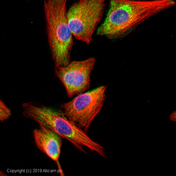
ab34151 stained in SKNSH cells. Cells were fixed with 100% methanol (5 min) at room temperature and incubated with PBS containing 10% goat serum, 0.3 M glycine, 1% BSA and 0.1% Triton for 1h at room temperature to permeabilise the cells and block non-specific protein-protein interactions. The cells were then incubated with the antibody ab34151 at 1µg/ml and ab7291 (Mouse monoclonal to alpha Tubulin - Loading Control) used at a 1/1000 dilution overnight at +4°C. The secondary antibodies were ab150081, Goat Anti-Rabbit IgG H&L (Alexa Fluor® 488) preadsorbed, (pseudo-colored green) and ab150120, Goat polyclonal Secondary Antibody to Mouse IgG - H&L (Alexa Fluor® 594) preadsorbed, (colored red), both used at a 1/1000 dilution for 1 hour at room temperature. DAPI was used to stain the cell nuclei (colored blue) at a concentration of 1.43 µM for 1hour at room temperature.
-
All lanes : Anti-Caspr antibody (ab34151) at 1/250 dilution
Lane 1 : Wild-type HAP1 whole cell lysate
Lane 2 : CNTNAP1 (Caspr) knockout HAP1 whole cell lysate
Lane 3 : MEF whole cell lysate
Lane 4 : HeLa whole cell lysate
Lysates/proteins at 20 µg per lane.
Predicted band size: 156 kDa
Observed band size: 180 kDa why is the actual band size different from the predicted?Lanes 1 - 4: Merged signal (red and green). Green - ab34151 observed at 180 kDa. Red - loading control, ab18058, observed at 130 kDa.
ab34151 was shown to recognize Caspr in wild-type HAP1 cells as signal was lost at the expected MW in CNTNAP1 (Caspr) knockout cells. Additional cross-reactive bands were observed in the wild-type and knockout cells. Wild-type and CNTNAP1 (Caspr) knockout samples were subjected to SDS-PAGE. ab34151 and ab18058 (Mouse anti-Vinculin loading control) were incubated overnight at 4°C at 1/250 dilution and 1/20000 dilution respectively. Blots were developed with Goat anti-Rabbit IgG H&L (IRDye® 800CW) preabsorbed ab216773 and Goat anti-Mouse IgG H&L (IRDye® 680RD) preabsorbed ab216776 secondary antibodies at 1/20000 dilution for 1 hour at room temperature before imaging.
-
Anti-Caspr antibody (ab34151) at 1/250 dilution + Brain (Rat) Whole Cell Lysate - normal tissue at 10 µg
Secondary
IRDye 680 Conjugated Goat Anti-Rabbit IgG (H+L) at 1/10000 dilution
Performed under reducing conditions.
Predicted band size: 156 kDa
Observed band size: 180 kDa why is the actual band size different from the predicted?
Additional bands at: 58 kDa. We are unsure as to the identity of these extra bands.
Caspr contains a number of potential glycosylation sites so it is thought that this is the reason it runs at 180kDa. -
Caspr was immunoprecipitated using 0.5mg Rat Brain whole tissue lysate, 5µg of Rabbit polyclonal to Caspr and 50µl of protein G magnetic beads (+). No antibody was added to the control (-).
The antibody was incubated under agitation with Protein G beads for 10min, Rat Brain whole tissue lysate lysate diluted in RIPA buffer was added to each sample and incubated for a further 10min under agitation.
Proteins were eluted by addition of 40µl SDS loading buffer and incubated for 10min at 70oC; 10µl of each sample was separated on a SDS PAGE gel, transferred to a nitrocellulose membrane, blocked with 5% BSA and probed with ab34151.
Secondary: Mouse monoclonal [SB62a] Secondary Antibody to Rabbit IgG light chain (HRP) (ab99697).
Band: 180kDa: Caspr.
Protocols
References (45)
ab34151 has been referenced in 45 publications.
- Tang SY et al. Caspr1 Facilitates sAPPa Production by Regulating a-Secretase ADAM9 in Brain Endothelial Cells. Front Mol Neurosci 13:23 (2020). PubMed: 32210761
- Fröb F et al. Ep400 deficiency in Schwann cells causes persistent expression of early developmental regulators and peripheral neuropathy. Nat Commun 10:2361 (2019). PubMed: 31142747
- Toomey LM et al. Comparing modes of delivery of a combination of ion channel inhibitors for limiting secondary degeneration following partial optic nerve transection. Sci Rep 9:15297 (2019). PubMed: 31653948
- Bonnefil V et al. Region-specific myelin differences define behavioral consequences of chronic social defeat stress in mice. Elife 8:N/A (2019). PubMed: 31407664
- Yermakov LM et al. Impairment of cognitive flexibility in type 2 diabetic db/db mice. Behav Brain Res 371:111978 (2019). PubMed: 31141724
Images
-
ab34151 stained in SKNSH cells. Cells were fixed with 100% methanol (5 min) at room temperature and incubated with PBS containing 10% goat serum, 0.3 M glycine, 1% BSA and 0.1% Triton for 1h at room temperature to permeabilise the cells and block non-specific protein-protein interactions. The cells were then incubated with the antibody ab34151 at 1µg/ml and ab7291 (Mouse monoclonal to alpha Tubulin - Loading Control) used at a 1/1000 dilution overnight at +4°C. The secondary antibodies were ab150081, Goat Anti-Rabbit IgG H&L (Alexa Fluor® 488) preadsorbed, (pseudo-colored green) and ab150120, Goat polyclonal Secondary Antibody to Mouse IgG - H&L (Alexa Fluor® 594) preadsorbed, (colored red), both used at a 1/1000 dilution for 1 hour at room temperature. DAPI was used to stain the cell nuclei (colored blue) at a concentration of 1.43 µM for 1hour at room temperature.
-
All lanes : Anti-Caspr antibody (ab34151) at 1/250 dilution
Lane 1 : Wild-type HAP1 whole cell lysate
Lane 2 : CNTNAP1 (Caspr) knockout HAP1 whole cell lysate
Lane 3 : MEF whole cell lysate
Lane 4 : HeLa whole cell lysate
Lysates/proteins at 20 µg per lane.
Predicted band size: 156 kDa
Observed band size: 180 kDa why is the actual band size different from the predicted?Lanes 1 - 4: Merged signal (red and green). Green - ab34151 observed at 180 kDa. Red - loading control, ab18058, observed at 130 kDa.
ab34151 was shown to recognize Caspr in wild-type HAP1 cells as signal was lost at the expected MW in CNTNAP1 (Caspr) knockout cells. Additional cross-reactive bands were observed in the wild-type and knockout cells. Wild-type and CNTNAP1 (Caspr) knockout samples were subjected to SDS-PAGE. ab34151 and ab18058 (Mouse anti-Vinculin loading control) were incubated overnight at 4°C at 1/250 dilution and 1/20000 dilution respectively. Blots were developed with Goat anti-Rabbit IgG H&L (IRDye® 800CW) preabsorbed ab216773 and Goat anti-Mouse IgG H&L (IRDye® 680RD) preabsorbed ab216776 secondary antibodies at 1/20000 dilution for 1 hour at room temperature before imaging.
-
Anti-Caspr antibody (ab34151) at 1/250 dilution + Brain (Rat) Whole Cell Lysate - normal tissue at 10 µg
Secondary
IRDye 680 Conjugated Goat Anti-Rabbit IgG (H+L) at 1/10000 dilution
Performed under reducing conditions.
Predicted band size: 156 kDa
Observed band size: 180 kDa why is the actual band size different from the predicted?
Additional bands at: 58 kDa. We are unsure as to the identity of these extra bands.
Caspr contains a number of potential glycosylation sites so it is thought that this is the reason it runs at 180kDa. -
Caspr was immunoprecipitated using 0.5mg Rat Brain whole tissue lysate, 5µg of Rabbit polyclonal to Caspr and 50µl of protein G magnetic beads (+). No antibody was added to the control (-).
The antibody was incubated under agitation with Protein G beads for 10min, Rat Brain whole tissue lysate lysate diluted in RIPA buffer was added to each sample and incubated for a further 10min under agitation.
Proteins were eluted by addition of 40µl SDS loading buffer and incubated for 10min at 70oC; 10µl of each sample was separated on a SDS PAGE gel, transferred to a nitrocellulose membrane, blocked with 5% BSA and probed with ab34151.
Secondary: Mouse monoclonal [SB62a] Secondary Antibody to Rabbit IgG light chain (HRP) (ab99697).
Band: 180kDa: Caspr.


