Anti-Brachyury / Bry antibody (ab20680)
Key features and details
- Rabbit polyclonal to Brachyury / Bry
- Suitable for: ICC/IF, IHC-P, WB
- Reacts with: Mouse, Human
- Isotype: IgG
Overview
-
Product name
Anti-Brachyury / Bry antibody
See all Brachyury / Bry primary antibodies -
Description
Rabbit polyclonal to Brachyury / Bry -
Host species
Rabbit -
Specificity
The immunogen used to raise this antibody has 81% homology with the Mouse Brachyury protein. Some customers have successfully used ab20680 on mouse samples, however we have not been successful detecting Brachyury in this species in our own testing and therefore cannot guarantee mouse reactivity. Please contact Abcam Scientific Support for more information.
-
Tested applications
Suitable for: ICC/IF, IHC-P, WBmore details -
Species reactivity
Reacts with: Mouse, Human
Predicted to work with: Dog Does not react with:
Zebrafish
 -
Immunogen
Synthetic peptide corresponding to Human Brachyury/ Bry aa 250-350 (internal sequence) conjugated to keyhole limpet haemocyanin.
(Peptide available asab21992)
Properties
-
Form
Liquid -
Storage instructions
Shipped at 4°C. Store at +4°C short term (1-2 weeks). Upon delivery aliquot. Store at -20°C or -80°C. Avoid freeze / thaw cycle. -
Storage buffer
pH: 7.40
Preservative: 0.02% Sodium azide
Constituent: PBS
Batches of this product that have a concentration Concentration information loading...
Purity
Immunogen affinity purifiedClonality
PolyclonalIsotype
IgGResearch areas
Associated products
-
Compatible Secondaries
-
Immunizing Peptide (Blocking)
-
Isotype control
-
Recombinant Protein
Applications
Our Abpromise guarantee covers the use of ab20680 in the following tested applications.
The application notes include recommended starting dilutions; optimal dilutions/concentrations should be determined by the end user.
Application Abreviews Notes ICC/IF Use at an assay dependent concentration. IHC-P Use at an assay dependent concentration. WB Use a concentration of 1 µg/ml. Detects a band of approximately 47 kDa (predicted molecular weight: 47 kDa). Target
-
Function
Involved in the transcriptional regulation of genes required for mesoderm formation and differentiation. Binds to a palindromic site (called T site) and activates gene transcription when bound to such a site. -
Involvement in disease
Genetic variations in T are associated with susceptibility to neural tube defects (NTD) [MIM:182940]. NTD are common congenital malformations. Spina bifida, which results from malformations in the caudal region of the neural tube, is compatible with life but associated with significant morbidity, including lower limb paralysis.
T is involved in susceptibility to the development of chordoma (CHDM) [MIM:215400]. Chordomas are rare, clinically malignant tumors derived from notochordal remnants. They occur along the length of the spinal axis, predominantly in the sphenooccipital, vertebral and sacrococcygeal regions. They are characterized by slow growth, local destruction of bone, extension into adjacent soft tissues and rarely, distant metastatic spread. Note=Susceptibility to development of chordomas is due to a T gene duplication. -
Sequence similarities
Contains 1 T-box DNA-binding domain. -
Cellular localization
Nucleus. - Information by UniProt
-
Database links
- Entrez Gene: 6862 Human
- Entrez Gene: 20997 Mouse
- Omim: 601397 Human
- SwissProt: O15178 Human
- SwissProt: P20293 Mouse
- Unigene: 389457 Human
- Unigene: 913 Mouse
-
Alternative names
- BRAC_HUMAN antibody
- Brachyury homolog antibody
- Brachyury protein antibody
see all
Images
-
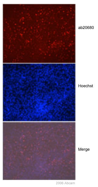
 Immunocytochemistry/ Immunofluorescence - Anti-Brachyury / Bry antibody (ab20680)This image is courtesy of Ludovic Valllier, University of Cambridge, UKAnti-Brachyury antibody, ab20680, stained nuclei of human embryonic stem cells differentiated into mesendoderm. As would be expected, mesoderm cells express Brachyury whereas endoderm cells are negative.
 -
 Immunohistochemistry (Formalin/PFA-fixed paraffin-embedded sections) - Anti-Brachyury / Bry antibody (ab20680)This image is courtesy of an Abreview submitted by Jim Manavisab20680 staining Brachyury in Human brain tissue sections by Immunohistochemistry (IHC-P - paraformaldehyde-fixed, paraffin-embedded sections). Tissue was fixed with formaldehyde and blocked with 3% serum for 30 minutes at room temperature; antigen retrieval was by heat mediation with a citrate buffer. Samples were incubated with primary antibody (1/2000 in horse serum) for 12 hours. A Streptavidin-conjugated Horse anti-rabbit monoclonal (1/250) was used as the secondary antibody.
 -
 Immunohistochemistry (Formalin/PFA-fixed paraffin-embedded sections) - Anti-Brachyury / Bry antibody (ab20680)This image is courtesy of Amanda Evans, University of CambridgeA proportion of nuclei from Day 3 Mouse EBs (derived from R1 cells) stained positive for Brachury using ab20680. In Day 8 EBs a vastly reduced proportion of nuclei stained positive (not shown). In day 10 EBs no nuclei stained Brachyury-positive. The IHC-P data for Brachyury in EBs corresponded with qRT-PCR expression data (not shown). Controls on Day 3 EBs in which primary or secondary antibody were omitted, undifferentiated ES cells and feeder cells (MEFs) all exhibited very little background staining. Zinc was used for tissue fixation.
 -
All lanes : Anti-Brachyury / Bry antibody (ab20680) at 1 µg/ml
Lane 1 : MUG-Chor1 (human sacral bone chordoma) at 10 µg
Lane 2 : MCF7 whole cell lysate (negative control) at 15 µg
Lane 3 : Human embryonic stem cells (pluripotent) (negative control) at 10 µg
Lane 4 : Human embryonic stem cells (mesoderm) at 10 µg
Secondary
All lanes : Goat polyclonal to Rabbit IgG - H&L - Pre-Adsorbed (HRP) at 1/50000 dilution
Developed using the ECL technique.
Performed under reducing conditions.
Predicted band size: 47 kDa
Observed band size: 50 kDa why is the actual band size different from the predicted?
Exposure time: 4 minutesThis blot was produced using a 4-12% Bis-tris gel under the MOPS buffer system. The gel was run at 200V for 50 minutes before being transferred onto a Nitrocellulose membrane at 30V for 70 minutes. The membrane was then blocked for an hour using 2% Bovine Serum Albumin before being incubated with ab20680 overnight at 4°C. Antibody binding was detected using an anti-rabbit antibody conjugated to HRP, and visualised using ECL development solution ab133406.
-
Anti-Brachyury / Bry antibody (ab20680) at 1 µg/ml + Whole Cell Lysate from Murine ES Cells Differentiated to Express Mesoderm Markers at 15 µg
Secondary
Goat polyclonal to Rabbit IgG - H&L - Pre-Adsorbed (HRP) at 1/3000 dilution
Performed under reducing conditions.
Predicted band size: 47 kDa
Observed band size: 53 kDa why is the actual band size different from the predicted?
Additional bands at: 75 kDa. We are unsure as to the identity of these extra bands.
ab20680 recognizes a band of 53 kDa in Murine Embryonic Stem Cells differentiated to express mesoderm markers. This corresponds in size to the Brachyury protein which has a predicted molecular weight of 47 kDa. A band of 75 kDa has also been observed, although we are unsure as to the identity of this band.
Protocols
References (41)
ab20680 has been referenced in 41 publications.
- Deuse T et al. Hypoimmunogenic derivatives of induced pluripotent stem cells evade immune rejection in fully immunocompetent allogeneic recipients. Nat Biotechnol 37:252-258 (2019). IHC-P ; Mouse . PubMed: 30778232
- Shrestha R et al. Aberrant hiPSCs-Derived from Human Keratinocytes Differentiates into 3D Retinal Organoids that Acquire Mature Photoreceptors. Cells 8:N/A (2019). IHC-Fr ; Human . PubMed: 30634512
- Ye B et al. LncKdm2b controls self-renewal of embryonic stem cells via activating expression of transcription factor Zbtb3. EMBO J 37:N/A (2018). PubMed: 29535137
- Gonzalez C et al. Modeling amyloid beta and tau pathology in human cerebral organoids. Mol Psychiatry 23:2363-2374 (2018). PubMed: 30171212
- Kumar D et al. Generation of three spinocerebellar ataxia type-12 patients derived induced pluripotent stem cell lines (IGIBi002-A, IGIBi003-A and IGIBi004-A). Stem Cell Res 31:216-221 (2018). ICC/IF ; Human . PubMed: 30130680
Images
-
 Immunocytochemistry/ Immunofluorescence - Anti-Brachyury / Bry antibody (ab20680) This image is courtesy of Ludovic Valllier, University of Cambridge, UKAnti-Brachyury antibody, ab20680, stained nuclei of human embryonic stem cells differentiated into mesendoderm. As would be expected, mesoderm cells express Brachyury whereas endoderm cells are negative.
 -
 Immunohistochemistry (Formalin/PFA-fixed paraffin-embedded sections) - Anti-Brachyury / Bry antibody (ab20680) This image is courtesy of an Abreview submitted by Jim Manavisab20680 staining Brachyury in Human brain tissue sections by Immunohistochemistry (IHC-P - paraformaldehyde-fixed, paraffin-embedded sections). Tissue was fixed with formaldehyde and blocked with 3% serum for 30 minutes at room temperature; antigen retrieval was by heat mediation with a citrate buffer. Samples were incubated with primary antibody (1/2000 in horse serum) for 12 hours. A Streptavidin-conjugated Horse anti-rabbit monoclonal (1/250) was used as the secondary antibody.
 -
 Immunohistochemistry (Formalin/PFA-fixed paraffin-embedded sections) - Anti-Brachyury / Bry antibody (ab20680) This image is courtesy of Amanda Evans, University of CambridgeA proportion of nuclei from Day 3 Mouse EBs (derived from R1 cells) stained positive for Brachury using ab20680. In Day 8 EBs a vastly reduced proportion of nuclei stained positive (not shown). In day 10 EBs no nuclei stained Brachyury-positive. The IHC-P data for Brachyury in EBs corresponded with qRT-PCR expression data (not shown). Controls on Day 3 EBs in which primary or secondary antibody were omitted, undifferentiated ES cells and feeder cells (MEFs) all exhibited very little background staining. Zinc was used for tissue fixation.
 -
All lanes : Anti-Brachyury / Bry antibody (ab20680) at 1 µg/ml
Lane 1 : MUG-Chor1 (human sacral bone chordoma) at 10 µg
Lane 2 : MCF7 whole cell lysate (negative control) at 15 µg
Lane 3 : Human embryonic stem cells (pluripotent) (negative control) at 10 µg
Lane 4 : Human embryonic stem cells (mesoderm) at 10 µg
Secondary
All lanes : Goat polyclonal to Rabbit IgG - H&L - Pre-Adsorbed (HRP) at 1/50000 dilution
Developed using the ECL technique.
Performed under reducing conditions.
Predicted band size: 47 kDa
Observed band size: 50 kDa why is the actual band size different from the predicted?
Exposure time: 4 minutesThis blot was produced using a 4-12% Bis-tris gel under the MOPS buffer system. The gel was run at 200V for 50 minutes before being transferred onto a Nitrocellulose membrane at 30V for 70 minutes. The membrane was then blocked for an hour using 2% Bovine Serum Albumin before being incubated with ab20680 overnight at 4°C. Antibody binding was detected using an anti-rabbit antibody conjugated to HRP, and visualised using ECL development solution ab133406.
-
Anti-Brachyury / Bry antibody (ab20680) at 1 µg/ml + Whole Cell Lysate from Murine ES Cells Differentiated to Express Mesoderm Markers at 15 µg
Secondary
Goat polyclonal to Rabbit IgG - H&L - Pre-Adsorbed (HRP) at 1/3000 dilution
Performed under reducing conditions.
Predicted band size: 47 kDa
Observed band size: 53 kDa why is the actual band size different from the predicted?
Additional bands at: 75 kDa. We are unsure as to the identity of these extra bands.
ab20680 recognizes a band of 53 kDa in Murine Embryonic Stem Cells differentiated to express mesoderm markers. This corresponds in size to the Brachyury protein which has a predicted molecular weight of 47 kDa. A band of 75 kDa has also been observed, although we are unsure as to the identity of this band.


