Anti-Bile Acid Receptor NR1H4 antibody (ab85606)
")
Key features and details
- Rabbit polyclonal to Bile Acid Receptor NR1H4
- Suitable for: ICC/IF, WB
- Reacts with: Human
- Isotype: IgG
Overview
-
Product name
Anti-Bile Acid Receptor NR1H4 antibody
See all Bile Acid Receptor NR1H4 primary antibodies -
Description
Rabbit polyclonal to Bile Acid Receptor NR1H4 -
Host species
Rabbit -
Tested Applications & Species
See all applications and species dataApplication Species ICC/IF HumanWB Human
-
Immunogen
Synthetic peptide corresponding to Bile Acid Receptor NR1H4 aa 50-150 conjugated to keyhole limpet haemocyanin.
(Peptide available asab97929) -
Positive control
- This antibody gave a positive signal in the following lysates: HepG2 Whole Cell; Human Kidney Tissue; Human Small Intestine.
Properties
-
Form
Liquid -
Storage instructions
Shipped at 4°C. Store at +4°C short term (1-2 weeks). Upon delivery aliquot. Store at -20°C or -80°C. Avoid freeze / thaw cycle. -
Storage buffer
pH: 7.40
Preservative: 0.02% Sodium azide
Constituent: PBS
Batches of this product that have a concentration Concentration information loading...
Purity
Immunogen affinity purifiedClonality
PolyclonalIsotype
IgGResearch areas
Associated products
-
Compatible Secondaries
-
Isotype control
-
Recombinant Protein
Applications
The Abpromise guarantee
Our Abpromise guarantee covers the use of ab85606 in the following tested applications.
The application notes include recommended starting dilutions; optimal dilutions/concentrations should be determined by the end user.
GuaranteedTested applications are guaranteed to work and covered by our Abpromise guarantee.
PredictedPredicted to work for this combination of applications and species but not guaranteed.
IncompatibleDoes not work for this combination of applications and species.
Application Species ICC/IF HumanWB HumanAll applications MouseHamsterCowApplication Abreviews Notes ICC/IF Use a concentration of 5 µg/ml.WB Use a concentration of 1 µg/ml. Detects a band of approximately 52 kDa (predicted molecular weight: 56 kDa).Notes ICC/IF
Use a concentration of 5 µg/ml.WB
Use a concentration of 1 µg/ml. Detects a band of approximately 52 kDa (predicted molecular weight: 56 kDa).Target
-
Function
Ligand-activated transcription factor. Receptor for bile acids such as chenodeoxycholic acid, lithocholic acid and deoxycholic acid. Represses the transcription of the cholesterol 7-alpha-hydroxylase gene (CYP7A1) through the induction of NR0B2 or FGF19 expression, via two distinct mechanisms. Activates the intestinal bile acid-binding protein (IBABP). Activates the transcription of bile salt export pump ABCB11 by directly recruiting histone methyltransferase CARM1 to this locus. -
Sequence similarities
Belongs to the nuclear hormone receptor family. NR1 subfamily.
Contains 1 nuclear receptor DNA-binding domain. -
Cellular localization
Nucleus. - Information by UniProt
-
Database links
- Entrez Gene: 9971 Human
- Entrez Gene: 20186 Mouse
- Omim: 603826 Human
- SwissProt: Q3SZL0 Cow
- SwissProt: Q96RI1 Human
- SwissProt: Q60641 Mouse
- Unigene: 282735 Human
- Unigene: 3095 Mouse
-
Alternative names
- BAR antibody
- Bile acid receptor antibody
- Farnesoid X activated receptor antibody
see all
Images
-
Western blot - Anti-Bile Acid Receptor NR1H4 antibody (ab85606)All lanes : Anti-Bile Acid Receptor NR1H4 antibody (ab85606) at 1 µg/ml
Lane 1 : HepG2 (Human hepatocellular liver carcinoma cell line) Whole Cell Lysate
Lane 2 : Human kidney tissue lysate - total protein (ab30203)
Lane 3 : Human small intestine tissue lysate - total protein (ab29276)
Lysates/proteins at 10 µg per lane.
Secondary
All lanes : Goat polyclonal to Rabbit IgG - H&L - Pre-Adsorbed (HRP) at 1/3000 dilution
Developed using the ECL technique.
Performed under reducing conditions.
Predicted band size: 56 kDa
Observed band size: 52 kDa why is the actual band size different from the predicted?
Exposure time: 20 minutes -
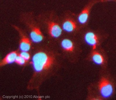 Immunocytochemistry/ Immunofluorescence - Anti-Bile Acid Receptor NR1H4 antibody (ab85606)ICC/IF image of ab85606 stained Hek293 cells. The cells were 100% methanol fixed (5 min) and then incubated in 1%BSA / 10% normal goat serum / 0.3M glycine in 0.1% PBS-Tween for 1h to permeabilise the cells and block non-specific protein-protein interactions. The cells were then incubated with the antibody (ab85606, 5µg/ml) overnight at +4°C. The secondary antibody (green) was Alexa Fluor® 488 goat anti-rabbit IgG (H+L) used at a 1/1000 dilution for 1h. Alexa Fluor® 594 WGA was used to label plasma membranes (red) at a 1/200 dilution for 1h. DAPI was used to stain the cell nuclei (blue) at a concentration of 1.43µM. This antibody also gave a positive result in 4% PFA fixed (10 min) HeLa and HepG2 cells at 5µg/ml.
Protocols
Datasheets and documents
References (0)
ab85606 has not yet been referenced specifically in any publications.
Images
-
Western blot - Anti-Bile Acid Receptor NR1H4 antibody (ab85606)All lanes : Anti-Bile Acid Receptor NR1H4 antibody (ab85606) at 1 µg/ml
Lane 1 : HepG2 (Human hepatocellular liver carcinoma cell line) Whole Cell Lysate
Lane 2 : Human kidney tissue lysate - total protein (ab30203)
Lane 3 : Human small intestine tissue lysate - total protein (ab29276)
Lysates/proteins at 10 µg per lane.
Secondary
All lanes : Goat polyclonal to Rabbit IgG - H&L - Pre-Adsorbed (HRP) at 1/3000 dilution
Developed using the ECL technique.
Performed under reducing conditions.
Predicted band size: 56 kDa
Observed band size: 52 kDa why is the actual band size different from the predicted?
Exposure time: 20 minutes
-
Immunocytochemistry/ Immunofluorescence - Anti-Bile Acid Receptor NR1H4 antibody (ab85606)ICC/IF image of ab85606 stained Hek293 cells. The cells were 100% methanol fixed (5 min) and then incubated in 1%BSA / 10% normal goat serum / 0.3M glycine in 0.1% PBS-Tween for 1h to permeabilise the cells and block non-specific protein-protein interactions. The cells were then incubated with the antibody (ab85606, 5µg/ml) overnight at +4°C. The secondary antibody (green) was Alexa Fluor® 488 goat anti-rabbit IgG (H+L) used at a 1/1000 dilution for 1h. Alexa Fluor® 594 WGA was used to label plasma membranes (red) at a 1/200 dilution for 1h. DAPI was used to stain the cell nuclei (blue) at a concentration of 1.43µM. This antibody also gave a positive result in 4% PFA fixed (10 min) HeLa and HepG2 cells at 5µg/ml.


