Anti-ATPB antibody - Mitochondrial Marker (ab128743)
")
Key features and details
- Rabbit polyclonal to ATPB - Mitochondrial Marker
- Suitable for: IHC-P, WB, ICC/IF
- Reacts with: Mouse, Rat, Human
- Isotype: IgG
Overview
-
Product name
Anti-ATPB antibody - Mitochondrial Marker
See all ATPB primary antibodies -
Description
Rabbit polyclonal to ATPB - Mitochondrial Marker -
Host species
Rabbit -
Tested applications
Suitable for: IHC-P, WB, ICC/IFmore details -
Species reactivity
Reacts with: Mouse, Rat, Human
Predicted to work with: Rabbit, Cow, Dog, Pig, Saccharomyces cerevisiae, Chimpanzee, Macaque monkey, Chinese hamster
-
Immunogen
Synthetic peptide corresponding to Human ATPB aa 150-250 conjugated to keyhole limpet haemocyanin.
(Peptide available asab140747) -
Positive control
- This antibody gave a positive signal in Mouse Liver tissue lysate as well as the following whole cell lysates: HeLa; HepG2; MEF1; NIH3T3; Raw264.7; PC12. This antibody gave a positive result in IHC in the following FFPE tissue: Human normal heart muscle. This antibody gave a positive result when used in the following methanol fixed cell lines: HepG2.
-
General notes
The Life Science industry has been in the grips of a reproducibility crisis for a number of years. Abcam is leading the way in addressing this with our range of recombinant monoclonal antibodies and knockout edited cell lines for gold-standard validation. Please check that this product meets your needs before purchasing.
If you have any questions, special requirements or concerns, please send us an inquiry and/or contact our Support team ahead of purchase. Recommended alternatives for this product can be found below, along with publications, customer reviews and Q&As
Images
-
Western blot - Anti-ATPB antibody - Mitochondrial Marker (ab128743)All lanes : Anti-ATPB antibody - Mitochondrial Marker (ab128743) at 1 µg/ml
Lane 1 : HeLa (Human epithelial carcinoma cell line) Whole Cell Lysate
Lane 2 : HepG2 (Human hepatocellular liver carcinoma cell line) Whole Cell Lysate
Lane 3 : MEF1 (Mouse embryonic fibroblast cell line) Whole Cell Lysate
Lane 4 : NIH 3T3 (Mouse embryonic fibroblast cell line) Whole Cell Lysate
Lane 5 : RAW 264.7 (Mouse leukaemic monocyte macrophage cell line) Whole Cell Lysate
Lane 6 : PC12 (Rat adrenal pheochromocytoma cell line) Whole Cell Lysate
Lane 7 : Liver (Mouse) Tissue Lysate
Lysates/proteins at 10 µg per lane.
Secondary
All lanes : Goat Anti-Rabbit IgG H&L (HRP) preadsorbed (ab97080) at 1/5000 dilution
Developed using the ECL technique.
Performed under reducing conditions.
Predicted band size: 56 kDa
Observed band size: 56 kDa
Exposure time: 10 seconds
This blot was produced using a 4-12% Bis-tris gel under the MOPS buffer system. The gel was run at 200V for 50 minutes before being transferred onto a Nitrocellulose membrane at 30V for 70 minutes. The membrane was then blocked for an hour using 5% Bovine Serum Albumin before being incubated with ab128743 overnight at 4°C. Antibody binding was detected using an anti-rabbit antibody conjugated to HRP, and visualised using ECL development solution. -
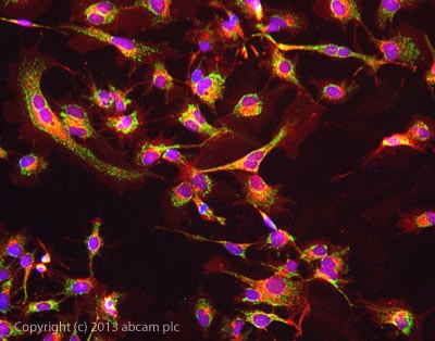 Immunocytochemistry/ Immunofluorescence - Anti-ATPB antibody - Mitochondrial Marker (ab128743)
ab128743 stained HepG2 cells. The cells were 100% methanol fixed (5 min) and then incubated in 1%BSA / 10% normal goat serum / 0.3M glycine in 0.1% PBS-Tween for 1h to permeabilise the cells and block non-specific protein-protein interactions. The cells were then incubated with the antibody ab128743 at 5µg/ml overnight at +4°C. The secondary antibody (green) was DyLight® 488 goat anti- rabbit (ab96899) IgG (H+L) used at a 1/250 dilution for 1h. Alexa Fluor® 594 WGA was used to label plasma membranes (red) at a 1/200 dilution for 1h. DAPI was used to stain the cell nuclei (blue) at a concentration of 1.43µM.
-
 Immunohistochemistry (Formalin/PFA-fixed paraffin-embedded sections) - Anti-ATPB antibody - Mitochondrial Marker (ab128743)
IHC image of ATPB staining in human normal heart muscle formalin fixed paraffin embedded tissue section, performed on a Leica BondTM system using the standard protocol F. The section was pre-treated using heat mediated antigen retrieval with sodium citrate buffer (pH6, epitope retrieval solution 1) for 20 mins. The section was then incubated with ab128743, 1µg/ml, for 15 mins at room temperature and detected using an HRP conjugated compact polymer system. DAB was used as the chromogen. The section was then counterstained with haematoxylin and mounted with DPX.
For other IHC staining systems (automated and non-automated) customers should optimize variable parameters such as antigen retrieval conditions, primary antibody concentration and antibody incubation times.
-
 Western blot - Anti-ATPB antibody - Mitochondrial Marker (ab128743) This image is courtesy of an anonymous AbreviewAll lanes : Anti-ATPB antibody - Mitochondrial Marker (ab128743) at 1/5000 dilution
Lane 1 : Saccharomyces cerevisiae strain BY4741 whole cell lysate
Lane 2 : Saccharomyces cerevisiae strain BY4741 delta ATP2 whole cell lysate
Lysates/proteins at 20 µg per lane.
Secondary
All lanes : HRP-conjugated goat anti-rabbit IgG monoclonal at 1/5000 dilution
Developed using the ECL technique.
Performed under reducing conditions.
Predicted band size: 56 kDa
Observed band size: 60 kDa why is the actual band size different from the predicted?
Additional bands at: 130 kDa (possible non-specific binding)
Exposure time: 5 minutes


