Anti-Aromatase antibody (ab35604)
Key features and details
- Rabbit polyclonal to Aromatase
- Suitable for: WB
- Reacts with: Human
- Isotype: IgG
Overview
-
Product name
Anti-Aromatase antibody
See all Aromatase primary antibodies -
Description
Rabbit polyclonal to Aromatase -
Host species
Rabbit -
Tested Applications & Species
See all applications and species dataApplication Species WB Human -
Immunogen
Synthetic peptide conjugated to KLH derived from within residues 450 to the C-terminus of Human Aromatase.
Read Abcam's proprietary immunogen policy (Peptide available as ab35661.)
Properties
-
Form
Liquid -
Storage instructions
Shipped at 4°C. Store at +4°C short term (1-2 weeks). Upon delivery aliquot. Store at -20°C or -80°C. Avoid freeze / thaw cycle. -
Storage buffer
pH: 7.40
Preservative: 0.02% Sodium azide
Constituent: PBS
Batches of this product that have a concentration Concentration information loading...
Purity
Immunogen affinity purifiedClonality
PolyclonalIsotype
IgGResearch areas
Associated products
-
Compatible Secondaries
-
Isotype control
-
Positive Controls
-
Recombinant Protein
Applications
The Abpromise guarantee
Our Abpromise guarantee covers the use of ab35604 in the following tested applications.
The application notes include recommended starting dilutions; optimal dilutions/concentrations should be determined by the end user.
GuaranteedTested applications are guaranteed to work and covered by our Abpromise guarantee.
PredictedPredicted to work for this combination of applications and species but not guaranteed.
IncompatibleDoes not work for this combination of applications and species.
Application Species WB HumanAll applications RabbitDogApplication Abreviews Notes WB (1) Use a concentration of 1 µg/ml. Detects a band of approximately 55 kDa (predicted molecular weight: 58 kDa).Notes WB
Use a concentration of 1 µg/ml. Detects a band of approximately 55 kDa (predicted molecular weight: 58 kDa).Target
-
Function
Catalyzes the formation of aromatic C18 estrogens from C19 androgens. -
Tissue specificity
Brain, placenta and gonads. -
Involvement in disease
Defects in CYP19A1 are a cause of aromatase excess syndrome (AEXS) [MIM:139300]; also known as familial gynecomastia. AEXS is characterized by an estrogen excess due to an increased aromatase activity.
Defects in CYP19A1 are the cause of aromatase deficiency (AROD) [MIM:107910]. AROD is a rare disease in which fetal androgens are not converted into estrogens due to placental aromatase deficiency. Thus, pregnant women exhibit a hirsutism, which spontaneously resolves after post-partum. At birth, female babies present with pseudohermaphroditism due to virilization of extern genital organs. In adult females, manifestations include delay of puberty, breast hypoplasia and primary amenorrhoea with multicystic ovaries. -
Sequence similarities
Belongs to the cytochrome P450 family. -
Cellular localization
Membrane. - Information by UniProt
-
Database links
- Entrez Gene: 1588 Human
- Omim: 107910 Human
- SwissProt: P11511 Human
- Unigene: 260074 Human
-
Alternative names
- ARO antibody
- ARO1 antibody
- Aromatase antibody
see all
Images
-
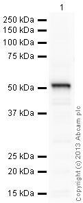
Anti-Aromatase antibody (ab35604) at 1 µg/ml + Human placenta tissue lysate - total protein (ab29745) at 20 µg
Secondary
Goat Anti-Rabbit IgG H&L (HRP) preadsorbed (ab97080) at 1/5000 dilution
Developed using the ECL technique.
Performed under reducing conditions.
Predicted band size: 58 kDa
Observed band size: 55 kDa why is the actual band size different from the predicted?
Exposure time: 150 secondsThis blot was produced using a 4-12% Bis-tris gel under the MOPS buffer system. The gel was run at 200V for 50 minutes before being transferred onto a Nitrocellulose membrane at 30V for 70 minutes. The membrane was then blocked for an hour using 5% Bovine Serum Albumin before being incubated with ab35604 overnight at 4°C. Antibody binding was detected using an anti-rabbit antibody conjugated to HRP, and visualised using ECL development solution.
-
Immunohistochemistry (Formalin/PFA-fixed paraffin-embedded sections) - Anti-Aromatase antibody (ab35604)Courtesy of Feng Y et al. Sci Rep. 2017; 7: 44810. doi: 10.1038/srep44810 Reproduced under the Creative Commons license http://creativecommons.org/licenses/by/4.0/.
Immunohistochemical analysis of adult proestrous mouse ovarian follicles staining CYP19/aromatase with ab35604, and VEGFA with ab46154. Staining of follicles at different stages using specific markers (upper panels) together with histological pictures using hematoxylin and eosin staining (lower panels).
Protocols
References (13)
ab35604 has been referenced in 13 publications.
- Zarate-Perez F et al. Biophysical characterization of Aptenodytes forsteri cytochrome P450 aromatase. J Inorg Biochem 184:79-87 (2018). WB . PubMed: 29684698
- Ke J et al. Prostaglandin E2 triggers cytochrome P450 17a hydroxylase overexpression via signal transducer and activator of transcription 3 phosphorylation and promotes invasion in endometrial cancer. Oncol Lett 16:4577-4585 (2018). PubMed: 30214592
- Adurthi S et al. Oestrogen Receptor-a binds the FOXP3 promoter and modulates regulatory T-cell function in human cervical cancer. Sci Rep 7:17289 (2017). IHC-P . PubMed: 29229929
- Cho S et al. Aromatase inhibitor regulates let-7 expression and let-7f-induced cell migration in endometrial cells from women with endometriosis. Fertil Steril 106:673-80 (2016). PubMed: 27320036
- Guven S et al. Functional maintenance of differentiated embryoid bodies in microfluidic systems: a platform for personalized medicine. Stem Cells Transl Med 4:261-8 (2015). ICC/IF ; Mouse . PubMed: 25666845
Images
-
Anti-Aromatase antibody (ab35604) at 1 µg/ml + Human placenta tissue lysate - total protein (ab29745) at 20 µg
Secondary
Goat Anti-Rabbit IgG H&L (HRP) preadsorbed (ab97080) at 1/5000 dilution
Developed using the ECL technique.
Performed under reducing conditions.
Predicted band size: 58 kDa
Observed band size: 55 kDa why is the actual band size different from the predicted?
Exposure time: 150 secondsThis blot was produced using a 4-12% Bis-tris gel under the MOPS buffer system. The gel was run at 200V for 50 minutes before being transferred onto a Nitrocellulose membrane at 30V for 70 minutes. The membrane was then blocked for an hour using 5% Bovine Serum Albumin before being incubated with ab35604 overnight at 4°C. Antibody binding was detected using an anti-rabbit antibody conjugated to HRP, and visualised using ECL development solution.
-
Immunohistochemistry (Formalin/PFA-fixed paraffin-embedded sections) - Anti-Aromatase antibody (ab35604) Courtesy of Feng Y et al. Sci Rep. 2017; 7: 44810. doi: 10.1038/srep44810 Reproduced under the Creative Commons license http://creativecommons.org/licenses/by/4.0/.
Immunohistochemical analysis of adult proestrous mouse ovarian follicles staining CYP19/aromatase with ab35604, and VEGFA with ab46154. Staining of follicles at different stages using specific markers (upper panels) together with histological pictures using hematoxylin and eosin staining (lower panels).


