Anti-Annexin-2/ANXA2 antibody (ab41803)
")
Key features and details
- Rabbit polyclonal to Annexin-2/ANXA2
- Suitable for: ICC/IF, WB, IHC-P
- Reacts with: Human
- Isotype: IgG
Overview
-
Product name
Anti-Annexin-2/ANXA2 antibody
See all Annexin-2/ANXA2 primary antibodies -
Description
Rabbit polyclonal to Annexin-2/ANXA2 -
Host species
Rabbit -
Tested Applications & Species
See all applications and species dataApplication Species ICC/IF HumanIHC-P HumanWB Human
-
Immunogen
Synthetic peptide corresponding to Human Annexin-2/ANXA2 aa 150-250 conjugated to keyhole limpet haemocyanin.
(Peptide available asab41802) -
Positive control
- WB: HeLa, HEK293, HepG2, MCF7 and SHSY-5Y whole cell lysates. ICC/IF: HeLa and MCF7 cells. IHC-P: Human breast carcinoma tissue.
-
General notes
This product was previously labelled as Annexin A2
Properties
-
Form
Liquid -
Storage instructions
Shipped at 4°C. Store at +4°C short term (1-2 weeks). Upon delivery aliquot. Store at -20°C or -80°C. Avoid freeze / thaw cycle. -
Storage buffer
pH: 7.40
Preservative: 0.02% Sodium azide
Constituent: PBS
Batches of this product that have a concentration Concentration information loading...
Purity
Immunogen affinity purifiedClonality
PolyclonalIsotype
IgGResearch areas
Associated products
-
Compatible Secondaries
-
Isotype control
-
Recombinant Protein
Applications
The Abpromise guarantee
Our Abpromise guarantee covers the use of ab41803 in the following tested applications.
The application notes include recommended starting dilutions; optimal dilutions/concentrations should be determined by the end user.
GuaranteedTested applications are guaranteed to work and covered by our Abpromise guarantee.
PredictedPredicted to work for this combination of applications and species but not guaranteed.
IncompatibleDoes not work for this combination of applications and species.
Application Species ICC/IF HumanIHC-P HumanWB HumanAll applications MouseRatSheepCowDogPigAfrican green monkeyApplication Abreviews Notes ICC/IF (5) Use a concentration of 1 µg/ml.WB (4) Use a concentration of 1 µg/ml. Detects a band of approximately 38 kDa (predicted molecular weight: 38 kDa).IHC-P (5) 1/2000. Perform heat mediated antigen retrieval before commencing with IHC staining protocol.Notes ICC/IF
Use a concentration of 1 µg/ml.WB
Use a concentration of 1 µg/ml. Detects a band of approximately 38 kDa (predicted molecular weight: 38 kDa).IHC-P
1/2000. Perform heat mediated antigen retrieval before commencing with IHC staining protocol.Target
-
Function
Calcium-regulated membrane-binding protein whose affinity for calcium is greatly enhanced by anionic phospholipids. It binds two calcium ions with high affinity. May be involved in heat-stress response. -
Sequence similarities
Belongs to the annexin family.
Contains 4 annexin repeats. -
Domain
A pair of annexin repeats may form one binding site for calcium and phospholipid. -
Post-translational
modificationsPhosphorylation of Tyr-24 enhances heat stress-induced translocation to the cell surface.
ISGylated. -
Cellular localization
Secreted > extracellular space > extracellular matrix > basement membrane. Melanosome. In the lamina beneath the plasma membrane. Identified by mass spectrometry in melanosome fractions from stage I to stage IV. Translocated from the cytoplasm to the cell surface through a Golgi-independent mechanism. - Information by UniProt
-
Database links
- Entrez Gene: 302 Human
- Entrez Gene: 12306 Mouse
- Entrez Gene: 56611 Rat
- Omim: 151740 Human
- SwissProt: P07355 Human
- SwissProt: P07356 Mouse
- SwissProt: Q07936 Rat
- Unigene: 511605 Human
see all -
Alternative names
- Annexin A2 antibody
- Annexin II antibody
- Annexin II, heavy chain antibody
see all
Images
-
Western blot - Anti-Annexin-2/ANXA2 antibody (ab41803)All lanes : Anti-Annexin-2/ANXA2 antibody (ab41803) at 1 µg/ml
Lane 1 : HeLa (Human epithelial carcinoma cell line) Whole Cell Lysate
Lane 2 : HEK293 (Human embryonic kidney cell line) Whole Cell Lysate
Lane 3 : HepG2 (Human hepatocellular liver carcinoma cell line) Whole Cell Lysate
Lane 4 : MCF7 (Human breast adenocarcinoma cell line) Whole Cell Lysate
Lane 5 : SHSY-5Y (Human neuroblastoma cell line) Whole Cell Lysate
Lysates/proteins at 10 µg per lane.
Secondary
All lanes : IRDye 680 Conjugated Goat Anti-Rabbit IgG (H+L) at 1/10000 dilution
Performed under reducing conditions.
Predicted band size: 38 kDa
Observed band size: 38 kDa -
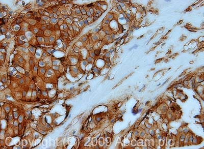 Immunohistochemistry (Formalin/PFA-fixed paraffin-embedded sections) - Anti-Annexin-2/ANXA2 antibody (ab41803)
IHC image of Annexin-2/ANXA2 staining in FFPE human breast carcinoma FFPE section, performed on a Leica BondTM system using the standard protocol F. The section was pre-treated using heat mediated antigen retrieval with sodium citrate buffer (pH6, epitope retrieval solution 1) for 20 mins. The section was then incubated with ab41803, 1µg/ml, for 15 mins at room temperature and detected using an HRP conjugated compact polymer system. DAB was used as the chromogen. The section was then counterstained with haematoxylin and mounted with DPX.
-
 Immunocytochemistry/ Immunofluorescence - Anti-Annexin-2/ANXA2 antibody (ab41803)ICC/IF image of ab41803 stained MCF7 cells. The cells were 100% methanol fixed (5 min) and then incubated in 1%BSA / 10% normal goat serum / 0.3M glycine in 0.1% PBS-Tween for 1h to permeabilise the cells and block non-specific protein-protein interactions. The cells were then incubated with the antibody (ab41803, 1µg/ml) overnight at +4°C. The secondary antibody (green) was Alexa Fluor® 488 goat anti-rabbit IgG (H+L) used at a 1/1000 dilution for 1h. Alexa Fluor® 594 WGA was used to label plasma membranes (red) at a 1/200 dilution for 1h. DAPI was used to stain the cell nuclei (blue) at a concentration of 1.43µM. This antibody also gave a positive result in 100% methanol fixed (5 min) HeLa, Hek293 and HepG2 cells at 1µg/ml, and in 4% PFA fixed (10 min) HeLa cells at 1µg/ml.
 -
 Immunocytochemistry/ Immunofluorescence - Anti-Annexin-2/ANXA2 antibody (ab41803)ICC/IF image of ab41803 stained HeLa cells. The cells were 100% methanol fixed (5 min) and then incubated in 1%BSA / 10% normal goat serum / 0.3M glycine in 0.1% PBS-Tween for 1h to permeabilise the cells and block non-specific protein-protein interactions. The cells were then incubated with the antibody (ab41803, 1µg/ml) overnight at +4°C. The secondary antibody (green) was DyLight® 488 goat anti-rabbit IgG - H&L, pre-adsorbed (ab96899) used at a 1/250 dilution for 1h. Alexa Fluor® 594 WGA was used to label plasma membranes (red) at a 1/200 dilution for 1h. DAPI was used to stain the cell nuclei (blue) at a concentration of 1.43µM.
 -
 Western blot - Anti-Annexin-2/ANXA2 antibody (ab41803)
Protocols
References (45)
ab41803 has been referenced in 45 publications.
- Agarwal A et al. Quantitative mass spectrometric analysis of the mouse cerebral cortex after ischemic stroke. PLoS One 15:e0231978 (2020). PubMed: 32315348
- Deng PC et al. LncRNA SNHG14 potentiates pancreatic cancer progression via modulation of annexin A2 expression by acting as a competing endogenous RNA for miR-613. J Cell Mol Med 23:7222-7232 (2019). PubMed: 31513352
- Xiong M et al. NUDT21 inhibits bladder cancer progression through ANXA2 and LIMK2 by alternative polyadenylation. Theranostics 9:7156-7167 (2019). PubMed: 31695759
- Nuerzhati Y et al. Role of the long non-coding RNA-Annexin A2 pseudogene 3/Annexin A2 signaling pathway in biliary atresia-associated hepatic injury. Int J Mol Med 43:739-748 (2019). PubMed: 30569159
- Luo GF et al. FOXD3 may be a new cellular target biomarker as a hypermethylation gene in human ovarian cancer. Cancer Cell Int 19:44 (2019). PubMed: 30858761
Images
-
Western blot - Anti-Annexin-2/ANXA2 antibody (ab41803)All lanes : Anti-Annexin-2/ANXA2 antibody (ab41803) at 1 µg/ml
Lane 1 : HeLa (Human epithelial carcinoma cell line) Whole Cell Lysate
Lane 2 : HEK293 (Human embryonic kidney cell line) Whole Cell Lysate
Lane 3 : HepG2 (Human hepatocellular liver carcinoma cell line) Whole Cell Lysate
Lane 4 : MCF7 (Human breast adenocarcinoma cell line) Whole Cell Lysate
Lane 5 : SHSY-5Y (Human neuroblastoma cell line) Whole Cell Lysate
Lysates/proteins at 10 µg per lane.
Secondary
All lanes : IRDye 680 Conjugated Goat Anti-Rabbit IgG (H+L) at 1/10000 dilution
Performed under reducing conditions.
Predicted band size: 38 kDa
Observed band size: 38 kDa
-
Immunohistochemistry (Formalin/PFA-fixed paraffin-embedded sections) - Anti-Annexin-2/ANXA2 antibody (ab41803)
IHC image of Annexin-2/ANXA2 staining in FFPE human breast carcinoma FFPE section, performed on a Leica BondTM system using the standard protocol F. The section was pre-treated using heat mediated antigen retrieval with sodium citrate buffer (pH6, epitope retrieval solution 1) for 20 mins. The section was then incubated with ab41803, 1µg/ml, for 15 mins at room temperature and detected using an HRP conjugated compact polymer system. DAB was used as the chromogen. The section was then counterstained with haematoxylin and mounted with DPX.
-
Immunocytochemistry/ Immunofluorescence - Anti-Annexin-2/ANXA2 antibody (ab41803)ICC/IF image of ab41803 stained MCF7 cells. The cells were 100% methanol fixed (5 min) and then incubated in 1%BSA / 10% normal goat serum / 0.3M glycine in 0.1% PBS-Tween for 1h to permeabilise the cells and block non-specific protein-protein interactions. The cells were then incubated with the antibody (ab41803, 1µg/ml) overnight at +4°C. The secondary antibody (green) was Alexa Fluor® 488 goat anti-rabbit IgG (H+L) used at a 1/1000 dilution for 1h. Alexa Fluor® 594 WGA was used to label plasma membranes (red) at a 1/200 dilution for 1h. DAPI was used to stain the cell nuclei (blue) at a concentration of 1.43µM. This antibody also gave a positive result in 100% methanol fixed (5 min) HeLa, Hek293 and HepG2 cells at 1µg/ml, and in 4% PFA fixed (10 min) HeLa cells at 1µg/ml.
-
Immunocytochemistry/ Immunofluorescence - Anti-Annexin-2/ANXA2 antibody (ab41803)ICC/IF image of ab41803 stained HeLa cells. The cells were 100% methanol fixed (5 min) and then incubated in 1%BSA / 10% normal goat serum / 0.3M glycine in 0.1% PBS-Tween for 1h to permeabilise the cells and block non-specific protein-protein interactions. The cells were then incubated with the antibody (ab41803, 1µg/ml) overnight at +4°C. The secondary antibody (green) was DyLight® 488 goat anti-rabbit IgG - H&L, pre-adsorbed (ab96899) used at a 1/250 dilution for 1h. Alexa Fluor® 594 WGA was used to label plasma membranes (red) at a 1/200 dilution for 1h. DAPI was used to stain the cell nuclei (blue) at a concentration of 1.43µM.
-
Western blot - Anti-Annexin-2/ANXA2 antibody (ab41803)


