Propidium Iodide (ab14083)
Overview
-
Product name
Propidium Iodide -
Product overview
Propidium Iodide (PI) binds to double-stranded DNA. PI cannot cross intact plasma membrane and therefore will only be present in DNA of cells where the plasma membrane has been compromised/ permeabilized.
For Microscopy analysis: PI can be viewed using rhodamine(red) filter (λ= 536/617). Cells will only be stained if the membrane has been permeated, either naturally (non-viable cells) or with detergents (for fluorescent staining).
For Flow Cytometry analysis: PI staining can be monitored in FL2 channel.
If used together as control for Annexin V assays ab14082, ab14083 or ab14152, PI should be diluted to 250 µg/ml solution (in PBS) prior use and added as 1 µl/ Annexin V Assay (0.25µg/assay).
Visit our FAQs page for tips and troubleshooting.
-
Tested applications
Suitable for: FM, Flow Cytmore details
Properties
-
Storage instructions
Store at +4°C. Please refer to protocols. -
Components Identifier 1 ml Propidium Iodide (1mg/ml) ab14083 1 x 1ml -
Research areas
-
Relevance
Propidium Iodide (PI) (MW=668.4 Da) is an intercalating agent and a fluorescent molecule which is membrane impermeant and generally excluded from viable cells. Upon entering cells, PI will bind to DNA and RNA by intercalating between bases. Once bound to the nucleic acids, its fluorescence is enhanced 20- to 30-fold. Exmax= 536nm / Emmax= 617 nm.
Images
-
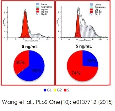
 Flow cytometry: ab14083 Image from Wang et al., PLoS One., 10(9). Fig 3a & c. doi:10.1371/journal.pone.0137712 Reproduced under the Creative Commons license http://creativecommons.org/licenses/by/4.0/
Wang et al. used ab14083 to investigate an optimized platform for maintaining proliferation of giant panda mesenchymal stem cells (MSCs). MSCs were fixed in 70% alcohol and 30% PBS at 4°C for 1 hour. Cells are then incubated in the dark with PBS, 20µg/ml propidium iodide (ab14083) and 1% RNaseA for 30 minutes at 37°C. Cell samples are resuspended in PBS and analyzed using FACS Calibur flow cytometry.
Flow cytometry analysis shows the cells are at different cell cycle stages in different concentrations (0ng/ml and 5ng/ml respectively) of basic fibroblast growth factor (bFGF).
-
 Immunofluorescence analysis using ab14083 Image from Melia MM et al ., Plos One.,29 (8). Fig 1b DOI: 10.1371/journal.pone.0106281. Reproduced under the Creative Commons license http://creativecommons.org/licenses/by/4.0/
Vero-DogSLAM (VDS) cells and Vero (African Green Monkey kidney) cells were infected with wtCDV. Cells were fixed, permeabilised and stained with SSPE serum and rabbit anti-human FITC; nuclei were stained using ab14083 propidium iodide. Images were taken using a Nikon Eclipse TE2000-U UV microscope (x400).


