Goat Anti-Mouse IgG H&L (DyLight® 488) preadsorbed (ab96879)
Key features and details
- Goat Anti-Mouse IgG H&L (DyLight® 488) preadsorbed
- Conjugation: DyLight® 488. Ex: 493nm, Em: 518nm
- Host species: Goat
- Isotype: IgG
- Suitable for: IHC-P, ICC/IF, Flow Cyt
Overview
-
Product name
Goat Anti-Mouse IgG H&L (DyLight® 488) preadsorbed
See all IgG secondary antibodies -
Host species
Goat -
Target species
Mouse -
Specificity
By immunoelectrophoresis and ELISA this antibody reacts specifically with Mouse IgG and with light chains common to other Mouse immunoglobulins. No antibody was detected against non immunoglobulin serum proteins. Reduced cross-reactivity to bovine, chicken, goat, horse, human, pig, rabbit and rat IgG was detected.
-
Tested applications
Suitable for: IHC-P, ICC/IF, Flow Cytmore details -
Minimal
cross-reactivity
Chicken, Cow, Goat, Horse, Human, Pig, Rabbit, Rat more details -
Conjugation
DyLight® 488. Ex: 493nm, Em: 518nm
Properties
-
Form
Liquid -
Storage instructions
Shipped at 4°C. Store at +4°C. -
Storage buffer
pH: 6.8
Preservative: 0.09% Sodium azide
Constituents: 0.2% BSA, PBS -
 Concentration information loading...
 -
Purity
Immunogen affinity purified -
Purification notes
Antiserum was cross adsorbed using bovine, chicken, horse, human, pig, rabbit and rat immunosorbents to remove cross reactive antibodies. This antibody was isolated by affinity chromatography using antigen coupled to agarose beads and conjugated to DyLight® 488. -
Clonality
Polyclonal -
Isotype
IgG -
Research areas
Images
-
 Flow Cytometry - Goat Anti-Mouse IgG H&L (DyLight® 488) preadsorbed (ab96879) This image is courtesy of an anonymous Abreview.
ab85137 staining S100 in a human melanoma cell line by Flow Cytometry. The cells were harvested using EDTA and washed in PBS. The sample was incubated with the primary antibody (1/100 in PBS) for 15 minutes at room temperature. A DyLight® 488-conjugated goat anti-mouse IgG H&L (ab96879) (1/100) was used as the secondary antibody.
-
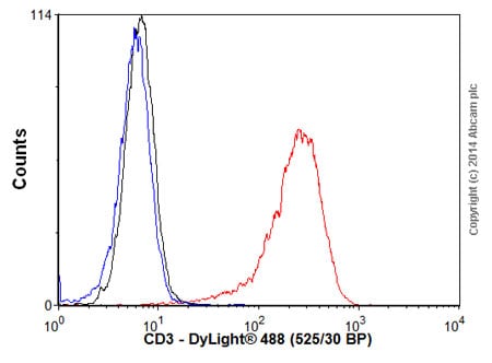
Overlay histogram showing Jurkat cells stained with ab8090 (red line). The cells were fixed with 4% paraformaldehyde (10 min) and then permeabilized with 0.1% PBS-Tween for 20 min. The cells were then incubated in 1x PBS / 10% normal goat serum / 0.3M glycine to block non-specific protein-protein interactions followed by the antibody (ab8090, 0.01μg/1x106 cells) for 30 min at 22°C. The secondary antibody Goat anti-mouse IgG H&L (DyLight® 488, pre-adsorbed) (ab96879) was used at 1/2000 dilution for 30 min at 22°C. Isotype control antibody (black line) was mouse IgG2a [ICIGG2A] (ab91361, 0.01μg/1x106 cells) used under the same conditions. Unlabelled sample (blue line) was also used as a control. Acquisition of >5,000 events were collected using a 20mW Argon ion laser (488nm) and 525/30 bandpass filter.
-
Overlay histogram showing HepG2 (Human liver hepatocellular carcinoma cell line) cells stained with ab2861 (red line).
The cells were fixed with 4% paraformaldehyde (10 min) and then permeabilized with 0.1% PBS-Tween for 20 min. The cells were then incubated in 1x PBS / 10% normal goat serum / 0.3M glycine to block non-specific protein-protein interactions followed by the antibody (ab2861, 1µg/1x106 cells) for 30 min at 22°C. The secondary antibody used was DyLight® 488 goat anti-mouse IgG (H+L) ab96879 at 1/500 dilution for 30 min at 22°C. Isotype control antibody (black line) was mouse IgG2a [ICIGG2A] (ab91361, 1µg/1x106 cells) used under the same conditions. Acquisition of >5,000 events was performed. -
Emission spectra of DyLight® fluorochromes available in our catalog.
Line colors represent the approximate visible colors of the wavelength maxima. -
 Immunocytochemistry/ Immunofluorescence - Goat Anti-Mouse IgG H&L (DyLight® 488) preadsorbed (ab96879)
ICC/IF image of ab40084 stained HepG2 cells. The cells were 4% PFA fixed (10 min) and then incubated in 1%BSA / 10% normal goat serum / 0.3M glycine in 0.1% PBS-Tween for 1h to permeabilise the cells and block non-specific protein-protein interactions. The cells were then incubated with the antibody (ab40084, 5µg/ml) overnight at +4°C. The secondary antibody (green) was DyLight® 488 goat anti-mouse IgG - H&L, pre-adsorbed (ab96879) used at a 1/250 dilution for 1h. Alexa Fluor® 594 WGA was used to label plasma membranes (red) at a 1/200 dilution for 1h. DAPI was used to stain the cell nuclei (blue) at a concentration of 1.43µM.


