Goat Anti-Mouse IgG H&L (HRP) (ab205719)
 (ab205719)")
Key features and details
- Goat Anti-Mouse IgG H&L (HRP)
- Conjugation: HRP
- Host species: Goat
- Isotype: IgG
- Suitable for: WB, IP, ELISA, IHC-P
Overview
-
Product name
Goat Anti-Mouse IgG H&L (HRP)
See all IgG secondary antibodies -
Host species
Goat -
Target species
Mouse -
Specificity
The antibody used for conjugation reacts with mouse immunoglobulins of all classes. Cross-reactions as determined by ELISA for the unconjugated antibody (ab182017): Chicken IgY, less than 2%. Human IgG, less than 6%. Rabbit IgG, less than 7%. Rat IgG, less than 47%. -
Tested applications
Suitable for: WB, IP, ELISA, IHC-Pmore details -
Immunogen
The details of the immunogen for this antibody are not available.
-
Conjugation
HRP
Properties
-
Form
Liquid -
Storage instructions
Shipped at 4°C. Store at +4°C short term (1-2 weeks). Upon delivery aliquot. Store at -20°C. Avoid freeze / thaw cycle. Store In the Dark. -
Storage buffer
pH: 7.40
Preservative: 0.1% Proclin 300 Solution
Constituents: PBS, 1% BSA, 30% Glycerol (glycerin, glycerine) -
 Concentration information loading...
 -
Purity
Immunogen affinity purified -
Purification notes
This antibody was isolated by affinity chromatography using antigen coupled to agarose beads and conjugated to Horse Radish Peroxidase (HRP). -
Clonality
Polyclonal -
Isotype
IgG -
Research areas
Images
-
Western blot - Goat Anti-Mouse IgG H&L (HRP) (ab205719)All lanes : Anti-alpha Tubulin antibody [DM1A] - Loading Control (ab7291) at 1 µg/ml
Lane 1 : Liver (Human) Tissue Lysate
Lane 2 : Liver (Mouse) Tissue Lysate
Lane 3 : Liver (Rat) Tissue Lysate
Lane 4 : HeLa (Human epithelial carcinoma cell line) Whole Cell Lysate
Lane 5 : NIH 3T3 (Mouse embryonic fibroblast cell line) Whole Cell Lysate
Lane 6 : PC12 (Rat adrenal pheochromocytoma cell line) Whole Cell Lysate
Lysates/proteins at 10 µg per lane.
Secondary
All lanes : Goat Anti-Mouse IgG H&L (HRP) (ab205719) at 1/5000 dilution
Developed using the ECL technique.
Performed under reducing conditions.
Observed band size: 52 kDa why is the actual band size different from the predicted?
Exposure time: 5 secondsThis blot was produced using a 4-12% Bis-tris gel under the MOPS buffer system. The gel was run at 200V for 50 minutes before being transferred onto a Nitrocellulose membrane at 30V for 70 minutes. The membrane was then blocked for an hour using 2% Bovine Serum Albumin before being incubated with ab7291 overnight at 4°C. Antibody binding was detected using ab205719, and visualised using ECL development solution ab133406.
-
 Immunohistochemistry (Formalin/PFA-fixed paraffin-embedded sections) - Goat Anti-Mouse IgG H&L (HRP) (ab205719)
IHC image of alpha tubulin staining in a section of formalin-fixed paraffin-embedded normal human colon tissue*. The section was pre-treated using pressure cooker heat mediated antigen retrieval with sodium citrate buffer (pH6) for 30mins, and incubated overnight at +4°C with ab7291 at 1/1000 dilution. An HRP-conjugated secondary (Ab205719, 1/10000 dilution) was used to detect the primary for 1hr at room temperature. DAB was used as the chromogen (ab103723), diluted 1/100 and incubated for 10min at room temperature. The section was counterstained with haematoxylin and mounted with DPX. The inset negative control image is taken from an identical assay without primary antibody.
For other IHC staining systems (automated and non-automated) customers should optimize variable parameters such as antigen retrieval conditions, primary antibody concentration and antibody incubation times.
*Tissue obtained from the Human Research Tissue Bank, supported by the NIHR Cambridge Biomedical Research Centre
-
 Western blot - Goat Anti-Mouse IgG H&L (HRP) (ab205719)All lanes : Anti-alpha Tubulin antibody [DM1A] - Loading Control (ab7291) at 1 µg/ml
Lane 1 : Liver (Human) Tissue Lysate
Lane 2 : Liver (Mouse) Tissue Lysate
Lane 3 : Liver (Rat) Tissue Lysate
Lysates/proteins at 10 µg per lane.
Secondary
All lanes : ab205719 (Left Image) at 1/5000 and a competitor secondary (Right Image) at 1/5000. Notice the decreased signal of the competitor product.
Performed under reducing conditions.
Observed band size: 52 kDa why is the actual band size different from the predicted?
Exposure time: 5 secondsThis blot was produced using a 4-12% Bis-tris gel under the MOPS buffer system. The gel was run at 200V for 50 minutes before being transferred onto a Nitrocellulose membrane at 30V for 70 minutes. The membrane was then blocked for an hour using 2% Bovine Serum Albumin before being incubated with ab7291 overnight at 4°C. Antibody binding was detected using ab205719 (Left Image) and a competitor secondary (Right Image), and visualised using ECL development solution ab133406.
-
 Immunohistochemistry (Formalin/PFA-fixed paraffin-embedded sections) - Goat Anti-Mouse IgG H&L (HRP) (ab205719)
IHC image of histone H4 staining in a section of formalin-fixed paraffin-embedded normal human colon tissue*. The section was pre-treated using pressure cooker heat mediated antigen retrieval with sodium citrate buffer (pH6) for 30mins, and incubated overnight at +4°C with ab31830 at 1/1000 dilution. An HRP-conjugated secondary (Ab205719, 1/10000 dilution) was used to detect the primary for 1hr at room temperature. DAB was used as the chromogen (ab103723), diluted 1/100 and incubated for 10min at room temperature. The section was counterstained with haematoxylin and mounted with DPX. The inset negative control image is taken from an identical assay without primary antibody.
For other IHC staining systems (automated and non-automated) customers should optimize variable parameters such as antigen retrieval conditions, primary antibody concentration and antibody incubation times.
*Tissue obtained from the Human Research Tissue Bank, supported by the NIHR Cambridge Biomedical Research Centre
-
 Western blot - Goat Anti-Mouse IgG H&L (HRP) (ab205719)All lanes : No Primary Antibody
Lane 1 : Liver (Human) Tissue Lysate
Lane 2 : Liver (Mouse) Tissue Lysate
Lane 3 : Liver (Rat) Tissue Lysate
Lysates/proteins at 10 µg per lane.
Secondary
All lanes : ab205719 (Left Image) 1/2000 and a competitor secondary (Right Image) 1/2000. Notice the increased background of the competitor product.
Performed under reducing conditions.
Exposure time: 10 secondsThis blot was produced using a 4-12% Bis-tris gel under the MOPS buffer system. The gel was run at 200V for 50 minutes before being transferred onto a Nitrocellulose membrane at 30V for 70 minutes. The membrane was incubated overnight with 2% Bovine Serum Albumin at 4°C. Any non-specific background binding was assessed by incubating the membrane with ab205719 (Left Image) and a competitor secondary (Right Image), and visualised using ECL development solution ab133406.
-
 Western blot - Goat Anti-Mouse IgG H&L (HRP) (ab205719)All lanes : Anti-beta Actin antibody [mAbcam 8226] - Loading Control (ab8226) at 1 µg/ml
Lane 1 : Liver (Human) Tissue Lysate
Lane 2 : Liver (Mouse) Tissue Lysate
Lane 3 : Liver (Rat) Tissue Lysate
Lane 4 : HeLa (Human epithelial carcinoma cell line) Whole Cell Lysate
Lane 5 : NIH 3T3 (Mouse embryonic fibroblast cell line) Whole Cell Lysate
Lane 6 : PC12 (Rat adrenal pheochromocytoma cell line) Whole Cell Lysate
Lysates/proteins at 10 µg per lane.
Secondary
All lanes : Goat Anti-Mouse IgG H&L (HRP) (ab205719) at 1/5000 dilution
Developed using the ECL technique.
Performed under reducing conditions.
Observed band size: 42 kDa why is the actual band size different from the predicted?
Exposure time: 10 secondsThis blot was produced using a 4-12% Bis-tris gel under the MOPS buffer system. The gel was run at 200V for 50 minutes before being transferred onto a Nitrocellulose membrane at 30V for 70 minutes. The membrane was then blocked for an hour using 2% Bovine Serum Albumin before being incubated with ab8226 overnight at 4°C. Antibody binding was detected using ab205719, and visualised using ECL development solution ab133406.
-
 ELISA - Goat Anti-Mouse IgG H&L (HRP) (ab205719)
Cross-reactivity of the polyclonal secondary antibody ab182017 was tested using a sandwich ELISA approach. The wells were coated with the indicated IgG standards at 1 µg/ml (50µl/well) and incubated overnight at 4°C, followed by a 5% BSA blocking step for 2h at RT. ab182017 was then added starting at 1 µg/ml and gradually diluted 1/4 (50 µl/well), followed by incubation for 2h. For the detection Donkey anti-Goat IgG H&L (HRP) (ab6885) was used at 1/10,000 dilution (50 µl/well), followed by incubation for 1h at RT.
Fot the batch tested, ab182017 showed a cross-reactivity below 2% towards Chicken IgY, 6% towards Human IgG, 7% towards Rabbit IgG and 47% towards Rat IgG.
This data was developed using the unconjugated antibody (ab182017).
-
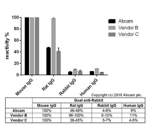 ELISA - Goat Anti-Mouse IgG H&L (HRP) (ab205719)
Cross-reactivity of Goat anti-Mouse IgG H&L (ab182017) and Goat anti-Mouse IgG H&L obtained from two different vendors was tested using a sandwich ELISA approach. The wells were coated with the indicated IgG standards (Rabbit, Human, Mouse and Rat) at 1 µg/ml (50µl/well) and incubated overnight at 4°C, followed by a 5% BSA blocking step for 2h at RT. Secondary antibodies were then added starting at 1 µg/ml and gradually diluted 1/4 (50 µl/well), followed by incubation for 2h. For the detection Donkey anti-Goat IgG H&L (HRP) (ab6885) was used at 1/10,000 dilution (50 µl/well), followed by incubation for 1h at RT. This data is from a representative dilution.
This data was developed using the unconjugated antibody (ab182017).


