Goat Anti-Mouse IgG H&L (Biotin) (ab207996)
 (ab207996)")
Key features and details
- Goat Anti-Mouse IgG H&L (Biotin)
- Conjugation: Biotin
- Host species: Goat
- Isotype: IgG
- Suitable for: Flow Cyt, WB, ICC/IF, IHC-Fr, IP, ELISA, IHC-P
Overview
-
Product name
Goat Anti-Mouse IgG H&L (Biotin)
See all IgG secondary antibodies -
Host species
Goat -
Target species
Mouse -
Specificity
The antibody used for conjugation reacts with mouse immunoglobulins of all classes. Cross-reactions as determined by ELISA for the unconjugated antibody (ab182017): Chicken IgY, less than 2%. Human IgG, less than 6%. Rabbit IgG, less than 7%. Rat IgG, less than 47%. -
Tested applications
Suitable for: Flow Cyt, WB, ICC/IF, IHC-Fr, IP, ELISA, IHC-Pmore details -
Immunogen
The details of the immunogen for this antibody are not available.
-
Conjugation
Biotin
Properties
-
Form
Liquid -
Storage instructions
Shipped at 4°C. Store at +4°C short term (1-2 weeks). Upon delivery aliquot. Store at -20°C. Avoid freeze / thaw cycle. Store In the Dark. -
Storage buffer
pH: 7.40
Preservative: 0.02% Sodium azide
Constituents: PBS, 1% BSA, 30% Glycerol (glycerin, glycerine) -
 Concentration information loading...
 -
Purity
Affinity purified -
Purification notes
Immunogen affinity purified - This antibody was isolated by affinity chromatography using antigen coupled to agarose beads and conjugated to Biotin. -
Clonality
Polyclonal -
Isotype
IgG -
Research areas
Images
-
Immunohistochemistry (Formalin/PFA-fixed paraffin-embedded sections) - Goat Anti-Mouse IgG H&L (Biotin) (ab207996)
IHC image of alpha Tubulin staining in a section of formalin-fixed paraffin-embedded normal human colon tissue*. Ab207996 Goat Anti-Mouse IgG H & L (Biotin) was used as the secondary antibody.
Staining was performed on a Leica BondTM. The section was pre-treated using heat mediated antigen retrieval with sodium citrate buffer (pH6, epitope retrieval solution 1) for 20 mins, before blocking of endogenous biotin using ab64212. The section was then incubated with ab7291, 1/100 dilution, for 15 mins at room temperature, followed by ab207996, 1/500 dilution, for 15 mins at room temperature. Detection was via an HRP conjugated ABC system and DAB was used as the chromogen. The section was then counterstained with haematoxylin and mounted with DPX.
The inset negative control image is taken from an identical assay without primary antibody.
For other IHC staining systems (automated and non-automated) customers should optimize variable parameters such as antigen retrieval conditions, primary antibody concentration and antibody incubation times.
*Tissue obtained from the Human Research Tissue Bank, supported by the NIHR Cambridge Biomedical Research Centre
-
 ELISA - Goat Anti-Mouse IgG H&L (Biotin) (ab207996)
ab207996 was tested by direct ELISA, where wells were coated with serially diluted mouse IgG (1000 – 16 ng/ml) for 2 hours, followed by a 2 hour blocking step (5% BSA). ab207996 (1:20,000 dilution; 2 hours) was added and detected by streptavidin-HRP (ab7403; 1:10,000 dilution; 1 hour). Signal was developed by TMB substrate. Data from duplicates; +/- SD.
-
 Immunohistochemistry (Formalin/PFA-fixed paraffin-embedded sections) - Goat Anti-Mouse IgG H&L (Biotin) (ab207996)
IHC image of Histone H4 staining in a section of formalin-fixed paraffin-embedded normal human colon tissue*. Ab207996 Goat Anti-Mouse IgG H & L (Biotin) was used as the secondary antibody.
Staining was performed on a Leica BondTM. The section was pre-treated using heat mediated antigen retrieval with sodium citrate buffer (pH6, epitope retrieval solution 1) for 20 mins, before blocking of endogenous biotin using ab64212. The section was then incubated with ab31830, 1/100 dilution, for 15 mins at room temperature, followed by ab207996, 1/500 dilution, for 15 mins at room temperature. Detection was via an HRP conjugated ABC system and DAB was used as the chromogen. The section was then counterstained with haematoxylin and mounted with DPX.
The inset negative control image is taken from an identical assay without primary antibody.
For other IHC staining systems (automated and non-automated) customers should optimize variable parameters such as antigen retrieval conditions, primary antibody concentration and antibody incubation times.
*Tissue obtained from the Human Research Tissue Bank, supported by the NIHR Cambridge Biomedical Research Centre
-
 ELISA - Goat Anti-Mouse IgG H&L (Biotin) (ab207996)
Cross-reactivity of the polyclonal secondary antibody ab182017 was tested using a sandwich ELISA approach. The wells were coated with the indicated IgG standards at 1 µg/ml (50µl/well) and incubated overnight at 4°C, followed by a 5% BSA blocking step for 2h at RT. ab182017 was then added starting at 1 µg/ml and gradually diluted 1/4 (50 µl/well), followed by incubation for 2h. For the detection Donkey anti-Goat IgG H&L (HRP) (ab6885) was used at 1/10,000 dilution (50 µl/well), followed by incubation for 1h at RT.
Fot the batch tested, ab182017 showed a cross-reactivity below 2% towards Chicken IgY, 6% towards Human IgG, 7% towards Rabbit IgG and 47% towards Rat IgG.
This data was developed using the unconjugated antibody (ab182017).
-
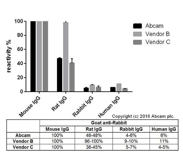 ELISA - Goat Anti-Mouse IgG H&L (Biotin) (ab207996)
Cross-reactivity of Goat anti-Mouse IgG H&L (ab182017) and Goat anti-Mouse IgG H&L obtained from two different vendors was tested using a sandwich ELISA approach. The wells were coated with the indicated IgG standards (Rabbit, Human, Mouse and Rat) at 1 µg/ml (50µl/well) and incubated overnight at 4°C, followed by a 5% BSA blocking step for 2h at RT. Secondary antibodies were then added starting at 1 µg/ml and gradually diluted 1/4 (50 µl/well), followed by incubation for 2h. For the detection Donkey anti-Goat IgG H&L (HRP) (ab6885) was used at 1/10,000 dilution (50 µl/well), followed by incubation for 1h at RT. This data is from a representative dilution.
This data was developed using the unconjugated antibody (ab182017).


