Anti-WASP/Wiskott-Aldrich syndrome protein antibody (ab74904)
Key features and details
- Rabbit polyclonal to WASP/Wiskott-Aldrich syndrome protein
- Suitable for: WB, IHC-P, ICC/IF
- Reacts with: Mouse, Human
- Isotype: IgG
Overview
-
Product name
Anti-WASP/Wiskott-Aldrich syndrome protein antibody
See all WASP/Wiskott-Aldrich syndrome protein primary antibodies -
Description
Rabbit polyclonal to WASP/Wiskott-Aldrich syndrome protein -
Host species
Rabbit -
Tested applications
Suitable for: WB, IHC-P, ICC/IFmore details -
Species reactivity
Reacts with: Mouse, Human -
Immunogen
Synthetic peptide corresponding to Human WASP/Wiskott-Aldrich syndrome protein aa 450 to the C-terminus conjugated to keyhole limpet haemocyanin.
(Peptide available asab74903) -
Positive control
- This antibody gave a positive signal in the following Whole Cell Lysates: Saos 2, LOVO, HT 1080, EBS E14 TG2A Day 0
-
General notes
This product was previously labelled as WASP
Reproducibility is key to advancing scientific discovery and accelerating scientists’ next breakthrough.
Abcam is leading the way with our range of recombinant antibodies, knockout-validated antibodies and knockout cell lines, all of which support improved reproducibility.
We are also planning to innovate the way in which we present recommended applications and species on our product datasheets, so that only applications & species that have been tested in our own labs, our suppliers or by selected trusted collaborators are covered by our Abpromise™ guarantee.
In preparation for this, we have started to update the applications & species that this product is Abpromise guaranteed for.
We are also updating the applications & species that this product has been “predicted to work with,” however this information is not covered by our Abpromise guarantee.
Applications & species from publications and Abreviews that have not been tested in our own labs or in those of our suppliers are not covered by the Abpromise guarantee.
Please check that this product meets your needs before purchasing. If you have any questions, special requirements or concerns, please send us an inquiry and/or contact our Support team ahead of purchase. Recommended alternatives for this product can be found below, as well as customer reviews and Q&As.
Properties
-
Form
Liquid -
Storage instructions
Shipped at 4°C. Store at +4°C short term (1-2 weeks). Upon delivery aliquot. Store at -20°C or -80°C. Avoid freeze / thaw cycle. -
Storage buffer
pH: 7.40
Preservative: 0.02% Sodium azide
Constituent: PBS
Batches of this product that have a concentration Concentration information loading...
Purity
Immunogen affinity purifiedClonality
PolyclonalIsotype
IgGResearch areas
Associated products
-
Compatible Secondaries
-
Isotype control
-
Recombinant Protein
Applications
Our Abpromise guarantee covers the use of ab74904 in the following tested applications.
The application notes include recommended starting dilutions; optimal dilutions/concentrations should be determined by the end user.
Application Abreviews Notes WB Use a concentration of 1 µg/ml. Detects a band of approximately 53 kDa (predicted molecular weight: 53 kDa). IHC-P Use a concentration of 5 µg/ml. ICC/IF Use a concentration of 5 µg/ml. Target
-
Function
Effector protein for Rho-type GTPases, providing a link with the Arp2/3 complex that regulates the structure and dynamics of the actin cytoskeleton. Important for efficient actin polymerization. Possible regulator of lymphocyte and platelet function. -
Tissue specificity
Expressed predominantly in the thymus. Also found, to a much lesser extent, in the spleen. -
Involvement in disease
Defects in WAS are the cause of Wiskott-Aldrich syndrome (WAS) [MIM:301000]; also known as eczema-thrombocytopenia-immunodeficiency syndrome. WAS is an X-linked recessive immunodeficiency characterized by eczema, thrombocytopenia, recurrent infections, and bloody diarrhea. Death usually occurs before age 10.
Defects in WAS are the cause of thrombocytopenia type 1 (THC1) [MIM:313900]. Thrombocytopenia is defined by a decrease in the number of platelets in circulating blood, resulting in the potential for increased bleeding and decreased ability for clotting.
Defects in WAS are a cause of neutropenia severe congenital X-linked (XLN) [MIM:300299]. XLN is an immunodeficiency syndrome characterized by recurrent major bacterial infections, severe congenital neutropenia, and monocytopenia. -
Sequence similarities
Contains 1 CRIB domain.
Contains 1 WH1 domain.
Contains 1 WH2 domain. -
Domain
The WH1 (Wasp homology 1) domain may bind a Pro-rich ligand.
The CRIB (Cdc42/Rac-interactive-binding) region binds to the C-terminal WH2 domain in the autoinhibited state of the protein. Binding of Rho-type GTPases to the CRIB induces a conformation change and leads to activation. -
Cellular localization
Cytoplasm > cytoskeleton. - Information by UniProt
-
Database links
- Entrez Gene: 7454 Human
- Entrez Gene: 22376 Mouse
- Omim: 300392 Human
- SwissProt: P42768 Human
- SwissProt: P70315 Mouse
- Unigene: 2157 Human
- Unigene: 4735 Mouse
-
Alternative names
- Eczema thrombocytopenia antibody
- IMD2 antibody
- SCNX antibody
see all
Images
-
All lanes : Anti-WASP/Wiskott-Aldrich syndrome protein antibody (ab74904) at 1 µg/ml
Lane 1 : Saos 2 (Human epithelial-like osteosarcoma cell line) Whole Cell Lysate
Lane 2 : LOVO (Human colon adenocarcinoma cell line) Whole Cell Lysate
Lane 3 : HT 1080 (Human fibrosarcoma) Whole Cell Lysate
Lane 4 : EBS E14 TG2A Day 0 (Mouse Pluripotent Embryonic Stem Cell) Whole Cell Lysate
Lysates/proteins at 10 µg per lane.
Secondary
All lanes : Goat polyclonal to Rabbit IgG - H&L - Pre-Adsorbed (HRP) at 1/3000 dilution
Developed using the ECL technique.
Performed under reducing conditions.
Predicted band size: 53 kDa
Observed band size: 53 kDa
Additional bands at: 100 kDa. We are unsure as to the identity of these extra bands.
Exposure time: 3 minutes -
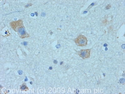
 Immunohistochemistry (Formalin/PFA-fixed paraffin-embedded sections) - Anti-WASP/Wiskott-Aldrich syndrome protein antibody (ab74904)
IHC image of WASP/Wiskott-Aldrich syndrome protein staining in human Cerebral cortex FFPE section, performed on a BondTM system using the standard protocol F. The section was pre-treated using heat mediated antigen retrieval with sodium citrate buffer (pH6, epitope retrieval solution 1) for 20 mins. The section was then incubated with ab74904, 5µg/ml, for 15 mins at room temperature and detected using an HRP conjugated compact polymer system. DAB was used as the chromogen. The section was then counterstained with haematoxylin and mounted with DPX
-
All lanes : Anti-WASP/Wiskott-Aldrich syndrome protein antibody (ab74904) at 1 µg/ml
Lane 1 : Saos-2
Lane 2 : LOVO (Human colon adenocarcinoma cell line) Whole Cell Lysate
Lane 3 : HT 1080 (Human fibrosarcoma) Whole Cell Lysate
Lane 4 : HepG2 (Human hepatocellular liver carcinoma cell line)
Lysates/proteins at 10 µg per lane.
Secondary
All lanes : Goat polyclonal to Rabbit IgG - H&L - Pre-Adsorbed (HRP) at 1/3000 dilution
Performed under reducing conditions.
Predicted band size: 53 kDa
Observed band size: 53 kDa
Additional bands at: 100 kDa. We are unsure as to the identity of these extra bands.
Exposure time: 20 minutes -
 Immunocytochemistry/ Immunofluorescence - Anti-WASP/Wiskott-Aldrich syndrome protein antibody (ab74904)ICC/IF image of ab74904 stained HepG2 cells. The cells were 4% PFA fixed (10 min) and then incubated in 1%BSA / 10% normal goat serum / 0.3M glycine in 0.1% PBS-Tween for 1h to permeabilise the cells and block non-specific protein-protein interactions. The cells were then incubated with the antibody (ab74904, 5µg/ml) overnight at +4°C. The secondary antibody (green) was ab96899 Dylight 488 goat anti-rabbit IgG (H+L) used at a 1/250 dilution for 1h. Alexa Fluor® 594 WGA was used to label plasma membranes (red) at a 1/200 dilution for 1h. DAPI was used to stain the cell nuclei (blue) at a concentration of 1.43µM.
Protocols
Datasheets and documents
References (1)
ab74904 has been referenced in 1 publication.
- Parisis N et al. Initiation of DNA replication requires actin dynamics and formin activity. EMBO J 36:3212-3231 (2017). PubMed: 28982779
Images
-
All lanes : Anti-WASP/Wiskott-Aldrich syndrome protein antibody (ab74904) at 1 µg/ml
Lane 1 : Saos 2 (Human epithelial-like osteosarcoma cell line) Whole Cell Lysate
Lane 2 : LOVO (Human colon adenocarcinoma cell line) Whole Cell Lysate
Lane 3 : HT 1080 (Human fibrosarcoma) Whole Cell Lysate
Lane 4 : EBS E14 TG2A Day 0 (Mouse Pluripotent Embryonic Stem Cell) Whole Cell Lysate
Lysates/proteins at 10 µg per lane.
Secondary
All lanes : Goat polyclonal to Rabbit IgG - H&L - Pre-Adsorbed (HRP) at 1/3000 dilution
Developed using the ECL technique.
Performed under reducing conditions.
Predicted band size: 53 kDa
Observed band size: 53 kDa
Additional bands at: 100 kDa. We are unsure as to the identity of these extra bands.
Exposure time: 3 minutes
-
 Immunohistochemistry (Formalin/PFA-fixed paraffin-embedded sections) - Anti-WASP/Wiskott-Aldrich syndrome protein antibody (ab74904)
IHC image of WASP/Wiskott-Aldrich syndrome protein staining in human Cerebral cortex FFPE section, performed on a BondTM system using the standard protocol F. The section was pre-treated using heat mediated antigen retrieval with sodium citrate buffer (pH6, epitope retrieval solution 1) for 20 mins. The section was then incubated with ab74904, 5µg/ml, for 15 mins at room temperature and detected using an HRP conjugated compact polymer system. DAB was used as the chromogen. The section was then counterstained with haematoxylin and mounted with DPX
-
All lanes : Anti-WASP/Wiskott-Aldrich syndrome protein antibody (ab74904) at 1 µg/ml
Lane 1 : Saos-2
Lane 2 : LOVO (Human colon adenocarcinoma cell line) Whole Cell Lysate
Lane 3 : HT 1080 (Human fibrosarcoma) Whole Cell Lysate
Lane 4 : HepG2 (Human hepatocellular liver carcinoma cell line)
Lysates/proteins at 10 µg per lane.
Secondary
All lanes : Goat polyclonal to Rabbit IgG - H&L - Pre-Adsorbed (HRP) at 1/3000 dilution
Performed under reducing conditions.
Predicted band size: 53 kDa
Observed band size: 53 kDa
Additional bands at: 100 kDa. We are unsure as to the identity of these extra bands.
Exposure time: 20 minutes
-
 Immunocytochemistry/ Immunofluorescence - Anti-WASP/Wiskott-Aldrich syndrome protein antibody (ab74904)ICC/IF image of ab74904 stained HepG2 cells. The cells were 4% PFA fixed (10 min) and then incubated in 1%BSA / 10% normal goat serum / 0.3M glycine in 0.1% PBS-Tween for 1h to permeabilise the cells and block non-specific protein-protein interactions. The cells were then incubated with the antibody (ab74904, 5µg/ml) overnight at +4°C. The secondary antibody (green) was ab96899 Dylight 488 goat anti-rabbit IgG (H+L) used at a 1/250 dilution for 1h. Alexa Fluor® 594 WGA was used to label plasma membranes (red) at a 1/200 dilution for 1h. DAPI was used to stain the cell nuclei (blue) at a concentration of 1.43µM.


