Anti-VPS35 antibody (ab118838)
")
Key features and details
- Rabbit polyclonal to VPS35
- Suitable for: WB, ICC/IF, IHC-P
- Knockout validated
- Reacts with: Human
- Isotype: IgG
Overview
-
Product name
Anti-VPS35 antibody
See all VPS35 primary antibodies -
Description
Rabbit polyclonal to VPS35 -
Host species
Rabbit -
Tested Applications & Species
See all applications and species dataApplication Species ICC/IF HumanIHC-P HumanWB Human
-
Immunogen
Synthetic peptide. This information is proprietary to Abcam and/or its suppliers.
-
Positive control
- This antibody gave a positive signal in the following whole cell lysates: HepG2; MOLT4; Raji. This antibody gave a positive result in IHC in the following FFPE tissue: Human normal heart muscle. This antibody gave a positive result when used in the following formaldehyde fixed cell lines: HepG2.
Properties
-
Form
Liquid -
Storage instructions
Shipped at 4°C. Store at +4°C short term (1-2 weeks). Upon delivery aliquot. Store at -20°C or -80°C. Avoid freeze / thaw cycle. -
Storage buffer
pH: 7.40
Preservative: 0.02% Sodium azide
Constituent: PBS
Batches of this product that have a concentration Concentration information loading...
Purity
Immunogen affinity purifiedClonality
PolyclonalIsotype
IgGResearch areas
Associated products
-
Compatible Secondaries
-
Isotype control
-
Recombinant Protein
Applications
The Abpromise guarantee
Our Abpromise guarantee covers the use of ab118838 in the following tested applications.
The application notes include recommended starting dilutions; optimal dilutions/concentrations should be determined by the end user.
GuaranteedTested applications are guaranteed to work and covered by our Abpromise guarantee.
PredictedPredicted to work for this combination of applications and species but not guaranteed.
IncompatibleDoes not work for this combination of applications and species.
Application Species ICC/IF HumanIHC-P HumanWB HumanAll applications MouseRatRabbitHorseCowDogPigChimpanzeeMacaque monkeyOrangutanApplication Abreviews Notes WB Use a concentration of 1 µg/ml. Detects a band of approximately 91 kDa (predicted molecular weight: 91 kDa).ICC/IF Use a concentration of 1 µg/ml.IHC-P Use a concentration of 5 µg/ml.Notes WB
Use a concentration of 1 µg/ml. Detects a band of approximately 91 kDa (predicted molecular weight: 91 kDa).ICC/IF
Use a concentration of 1 µg/ml.IHC-P
Use a concentration of 5 µg/ml.Target
-
Function
Essential component of the retromer complex, a complex required to retrieve lysosomal enzyme receptors (IGF2R and M6PR) from endosomes to the trans-Golgi network. Also required to regulate transcytosis of the polymeric immunoglobulin receptor (pIgR-pIgA). -
Tissue specificity
Ubiquitous. Highly expressed in heart, brain, placenta, skeletal muscle, spleen, thymus, testis, ovary, small intestine, kidney and colon. -
Sequence similarities
Belongs to the VPS35 family. -
Cellular localization
Cytoplasm. Membrane. - Information by UniProt
-
Database links
- Entrez Gene: ID: 465139 Chimpanzee
- Entrez Gene: 521864 Cow
- Entrez Gene: 100056512 Horse
- Entrez Gene: 100133770 Human
- Entrez Gene: 55737 Human
- Entrez Gene: 65114 Mouse
- Entrez Gene: 100174173 Orangutan
- Entrez Gene: 100623578 Pig
see all -
Alternative names
- DKFZp434E1211 antibody
- DKFZp434P1672 antibody
- FLJ10752 antibody
see all
Images
-
Western blot - Anti-VPS35 antibody (ab118838)
Lane 1: Wild-type HAP1 cell lysate (20 µg)
Lane 2: VPS35 knockout HAP1 cell lysate (20 µg)
Lane 3: NIH/3T3 cell lysate (20 µg)
Lane 4: A549 cell lysate (20 µg)
Lanes 1 - 4: Merged signal (red and green). Green - ab118838 observed at 90 kDa. Red - loading control, ab8245, observed at 37 kDa.
ab118838 was shown to recognize VPS35 in wild-type HAP1 cells along with additional cross-reactive bands. No band was observed when VPS35 knockout samples were examined. Wild-type and VPS35 knockout samples were subjected to SDS-PAGE. ab118838 and ab8245 (loading control to GAPDH) were diluted 1μg/mL and 1/2000 and incubated overnight at 4°C. Blots were developed with Goat anti-Rabbit IgG H&L (IRDye® 800CW) preadsorbed ab216773 and Goat anti-Mouse IgG H&L (IRDye® 680RD) preadsorbed ab216776 secondary antibodies at 1/10,000 dilution for 1 hour at room temperature before imaging. -
 Immunocytochemistry/ Immunofluorescence - Anti-VPS35 antibody (ab118838)
ICC/IF image of ab118838 stained HepG2 cells. The cells were 4% formaldehyde fixed (10 min) and then incubated in 1%BSA / 10% normal goat serum / 0.3M glycine in 0.1% PBS-Tween for 1h to permeabilise the cells and block non-specific protein-protein interactions. The cells were then incubated with the antibody ab118838 at 1µg/ml overnight at +4°C. The secondary antibody (green) was DyLight® 488 goat anti- rabbit (ab96899) IgG (H+L) used at a 1/250 dilution for 1h. Alexa Fluor® 594 WGA was used to label plasma membranes (red) at a 1/200 dilution for 1h. DAPI was used to stain the cell nuclei (blue) at a concentration of 1.43µM.
-
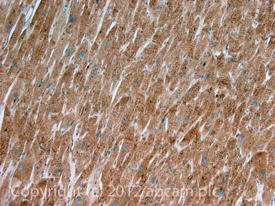 Immunohistochemistry (Formalin/PFA-fixed paraffin-embedded sections) - Anti-VPS35 antibody (ab118838)
IHC image of VPS35 staining in Human normal heart muscle formalin fixed paraffin embedded tissue section, performed on a Leica BondTM system using the standard protocol F. The section was pre-treated using heat mediated antigen retrieval with sodium citrate buffer (pH6, epitope retrieval solution 1) for 20 mins. The section was then incubated with ab118838, 5µg/ml, for 15 mins at room temperature and detected using an HRP conjugated compact polymer system. DAB was used as the chromogen. The section was then counterstained with haematoxylin and mounted with DPX.
For other IHC staining systems (automated and non-automated) customers should optimize variable parameters such as antigen retrieval conditions, primary antibody concentration and antibody incubation times.
-
 Western blot - Anti-VPS35 antibody (ab118838)All lanes : Anti-VPS35 antibody (ab118838) at 1 µg/ml
Lane 1 : HepG2 (Human hepatocellular liver carcinoma cell line) Whole Cell Lysate
Lane 2 : MOLT4 (Human acute lymphoblastic leukemia cell line) Whole Cell Lysate
Lane 3 : Raji (Human Burkitt's lymphoma cell line) Whole Cell Lysate
Lysates/proteins at 10 µg per lane.
Secondary
All lanes : Goat Anti-Rabbit IgG H&L (HRP) preadsorbed (ab97080) at 1/5000 dilution
Developed using the ECL technique.
Performed under reducing conditions.
Predicted band size: 91 kDa
Observed band size: 85 kDa why is the actual band size different from the predicted?
Additional bands at: 24 kDa, 35 kDa, 71 kDa. We are unsure as to the identity of these extra bands.
Exposure time: 2 minutes
Protocols
Datasheets and documents
References (0)
ab118838 has not yet been referenced specifically in any publications.
Images
-
Western blot - Anti-VPS35 antibody (ab118838)
Lane 1: Wild-type HAP1 cell lysate (20 µg)
Lane 2: VPS35 knockout HAP1 cell lysate (20 µg)
Lane 3: NIH/3T3 cell lysate (20 µg)
Lane 4: A549 cell lysate (20 µg)
Lanes 1 - 4: Merged signal (red and green). Green - ab118838 observed at 90 kDa. Red - loading control, ab8245, observed at 37 kDa.
ab118838 was shown to recognize VPS35 in wild-type HAP1 cells along with additional cross-reactive bands. No band was observed when VPS35 knockout samples were examined. Wild-type and VPS35 knockout samples were subjected to SDS-PAGE. ab118838 and ab8245 (loading control to GAPDH) were diluted 1μg/mL and 1/2000 and incubated overnight at 4°C. Blots were developed with Goat anti-Rabbit IgG H&L (IRDye® 800CW) preadsorbed ab216773 and Goat anti-Mouse IgG H&L (IRDye® 680RD) preadsorbed ab216776 secondary antibodies at 1/10,000 dilution for 1 hour at room temperature before imaging. -
Immunocytochemistry/ Immunofluorescence - Anti-VPS35 antibody (ab118838)
ICC/IF image of ab118838 stained HepG2 cells. The cells were 4% formaldehyde fixed (10 min) and then incubated in 1%BSA / 10% normal goat serum / 0.3M glycine in 0.1% PBS-Tween for 1h to permeabilise the cells and block non-specific protein-protein interactions. The cells were then incubated with the antibody ab118838 at 1µg/ml overnight at +4°C. The secondary antibody (green) was DyLight® 488 goat anti- rabbit (ab96899) IgG (H+L) used at a 1/250 dilution for 1h. Alexa Fluor® 594 WGA was used to label plasma membranes (red) at a 1/200 dilution for 1h. DAPI was used to stain the cell nuclei (blue) at a concentration of 1.43µM.
-
Immunohistochemistry (Formalin/PFA-fixed paraffin-embedded sections) - Anti-VPS35 antibody (ab118838)
IHC image of VPS35 staining in Human normal heart muscle formalin fixed paraffin embedded tissue section, performed on a Leica BondTM system using the standard protocol F. The section was pre-treated using heat mediated antigen retrieval with sodium citrate buffer (pH6, epitope retrieval solution 1) for 20 mins. The section was then incubated with ab118838, 5µg/ml, for 15 mins at room temperature and detected using an HRP conjugated compact polymer system. DAB was used as the chromogen. The section was then counterstained with haematoxylin and mounted with DPX.
For other IHC staining systems (automated and non-automated) customers should optimize variable parameters such as antigen retrieval conditions, primary antibody concentration and antibody incubation times.
-
Western blot - Anti-VPS35 antibody (ab118838)All lanes : Anti-VPS35 antibody (ab118838) at 1 µg/ml
Lane 1 : HepG2 (Human hepatocellular liver carcinoma cell line) Whole Cell Lysate
Lane 2 : MOLT4 (Human acute lymphoblastic leukemia cell line) Whole Cell Lysate
Lane 3 : Raji (Human Burkitt's lymphoma cell line) Whole Cell Lysate
Lysates/proteins at 10 µg per lane.
Secondary
All lanes : Goat Anti-Rabbit IgG H&L (HRP) preadsorbed (ab97080) at 1/5000 dilution
Developed using the ECL technique.
Performed under reducing conditions.
Predicted band size: 91 kDa
Observed band size: 85 kDa why is the actual band size different from the predicted?
Additional bands at: 24 kDa, 35 kDa, 71 kDa. We are unsure as to the identity of these extra bands.
Exposure time: 2 minutes


