Anti-TLE3/ESG antibody (ab94972)
Key features and details
- Rabbit polyclonal to TLE3/ESG
- Suitable for: IP, WB, ICC/IF
- Reacts with: Human
- Isotype: IgG
Overview
-
Product name
Anti-TLE3/ESG antibody
See all TLE3/ESG primary antibodies -
Description
Rabbit polyclonal to TLE3/ESG -
Host species
Rabbit -
Tested applications
Suitable for: IP, WB, ICC/IFmore details -
Species reactivity
Reacts with: Human
Predicted to work with: Mouse, Rat
-
Immunogen
Synthetic peptide. This information is proprietary to Abcam and/or its suppliers.
-
Positive control
- This antibody gave a positive signal in the following lysates: Breast (Human) Tissue Lysate; MCF7 Whole Cell Lysate; MDA-MB-231 Whole Cell Lysate; Lung (Human) Tissue Lysate; A549 Whole Cell Lysate; A431 Whole Cell Lysate; HeLa Nuclear Lysate
-
General notes
The Life Science industry has been in the grips of a reproducibility crisis for a number of years. Abcam is leading the way in addressing this with our range of recombinant monoclonal antibodies and knockout edited cell lines for gold-standard validation. Please check that this product meets your needs before purchasing.
If you have any questions, special requirements or concerns, please send us an inquiry and/or contact our Support team ahead of purchase. Recommended alternatives for this product can be found below, along with publications, customer reviews and Q&As
Properties
-
Form
Liquid -
Storage instructions
Shipped at 4°C. Store at +4°C short term (1-2 weeks). Upon delivery aliquot. Store at -20°C or -80°C. Avoid freeze / thaw cycle. -
Storage buffer
pH: 7.40
Preservative: 0.02% Sodium azide
Constituent: PBS
Batches of this product that have a concentration Concentration information loading...
Purity
Immunogen affinity purifiedClonality
PolyclonalIsotype
IgGResearch areas
Associated products
-
Compatible Secondaries
-
Isotype control
Applications
The Abpromise guarantee
Our Abpromise guarantee covers the use of ab94972 in the following tested applications.
The application notes include recommended starting dilutions; optimal dilutions/concentrations should be determined by the end user.
Application Abreviews Notes IP (1) Use at an assay dependent concentration.WB (1) Use a concentration of 1 µg/ml. Detects a band of approximately 83 kDa (predicted molecular weight: 83 kDa).ICC/IF (1) Use a concentration of 1 µg/ml.Notes IP
Use at an assay dependent concentration.WB
Use a concentration of 1 µg/ml. Detects a band of approximately 83 kDa (predicted molecular weight: 83 kDa).ICC/IF
Use a concentration of 1 µg/ml.Target
-
Function
Transcriptional corepressor that binds to a number of transcription factors. Inhibits the transcriptional activation mediated by CTNNB1 and TCF family members in Wnt signaling. The effects of full-length TLE family members may be modulated by association with dominant-negative AES. -
Tissue specificity
Placenta and lung. -
Sequence similarities
Belongs to the WD repeat Groucho/TLE family.
Contains 7 WD repeats. -
Post-translational
modificationsUbiquitinated by XIAP/BIRC4. This ubiquitination does not affect its stability, nuclear localization, or capacity to tetramerize but inhibits its interaction with TCF7L2/TCF4. -
Cellular localization
Nucleus. - Information by UniProt
-
Database links
- Entrez Gene: 7090 Human
- Entrez Gene: 21887 Mouse
- Entrez Gene: 84424 Rat
- Omim: 600190 Human
- SwissProt: Q04726 Human
- SwissProt: Q08122 Mouse
- SwissProt: Q9JIT3 Rat
- Unigene: 287362 Human
see all -
Alternative names
- E(sp1) homolog Drosophila antibody
- Enhancer of split groucho 3 antibody
- Enhancer of split groucho-like protein 3 antibody
see all
Images
-
ab94972 staining Transducin-like enhancer protein 3 in HeLa cells. The cells were fixed with 4% paraformaldehyde (10 min), permeabilized with 0.1% PBS-Triton X-100 for 5 minutes and then blocked with 1% BSA/10% normal goat serum/0.3M glycine in 0.1% PBS-Tween for 1h. The cells were then incubated overnight at 4°C with ab94972 at 0.1µg/ml and ab7291, Mouse monoclonal [DM1A] to alpha Tubulin - Loading Control. Cells were then incubated with ab150081, Goat polyclonal Secondary Antibody to Rabbit IgG - H&L (Alexa Fluor® 488), pre-adsorbed at 1/1000 dilution (shown in green) and ab150120, Goat polyclonal Secondary Antibody to Mouse IgG - H&L (Alexa Fluor® 594), pre-adsorbed at 1/1000 dilution (shown in pseudocolour red). Nuclear DNA was labelled with DAPI (shown in blue).
Also suitable in cells fixed with 100% methanol (5 min).
Image was acquired with a high-content analyser (Operetta CLS, Perkin Elmer) and a maximum intensity projection of confocal sections is shown.
-
All lanes : Anti-TLE3/ESG antibody (ab94972) at 1 µg/ml
Lane 1 : Human breast tissue lysate - total protein (ab30090)
Lane 2 : MCF7 (Human breast adenocarcinoma cell line) Whole Cell Lysate
Lane 3 : MDA-MB-231 (Human breast adenocarcinoma cell line) Whole Cell Lysate
Lane 4 : Lung (Human) Tissue Lysate
Lane 5 : A549 (Human lung adenocarcinoma epithelial cell line) Whole Cell Lysate
Lane 6 : A431 (Human epithelial carcinoma cell line) Whole Cell Lysate
Lane 7 : HeLa (Human epithelial carcinoma cell line) Nuclear Lysate
Lysates/proteins at 10 µg per lane.
Secondary
All lanes : Goat polyclonal Secondary Antibody to Rabbit IgG - H&L (HRP), pre-adsorbed at 1/5000 dilution
Developed using the ECL technique.
Performed under reducing conditions.
Predicted band size: 83 kDa
Observed band size: 83 kDa
Additional bands at: 125 kDa, 43 kDa. We are unsure as to the identity of these extra bands.
Exposure time: 8 minutes
Abcam recommends using milk as the blocking agent. Abcam welcomes customer feedback and would appreciate any comments regarding this product and the data presented above. -
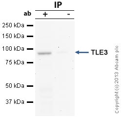
TLE3/ESG was immunoprecipitated using 0.5mg HeLa whole cell extract, 5µg of Rabbit polyclonal to TLE3/ESG and 50µl of protein G magnetic beads (+). No antibody was added to the control (-).
The antibody was incubated under agitation with Protein G beads for 10min, HeLa whole cell extract lysate diluted in RIPA buffer was added to each sample and incubated for a further 10min under agitation.
Proteins were eluted by addition of 40µl SDS loading buffer and incubated for 10min at 70°C; 10µl of each sample was separated on a SDS PAGE gel, transferred to a nitrocellulose membrane, blocked with 5% BSA and probed with ab94972.
Secondary: Mouse monoclonal [SB62a] Secondary Antibody to Rabbit IgG light chain (HRP) (ab99697).
Band: 83kDa; TLE3/ESG
Protocols
Datasheets and documents
-
SDS download
-
Datasheet download
References (3)
ab94972 has been referenced in 3 publications.
- Simeoni F et al. Enhancer recruitment of transcription repressors RUNX1 and TLE3 by mis-expressed FOXC1 blocks differentiation in acute myeloid leukemia. Cell Rep 36:109725 (2021). PubMed: 34551306
- Yang RW et al. TLE3 represses colorectal cancer proliferation by inhibiting MAPK and AKT signaling pathways. J Exp Clin Cancer Res 35:152 (2016). PubMed: 27669982
- Bara AM et al. Generation of a TLE3 heterozygous knockout human embryonic stem cell line using CRISPR-Cas9. Stem Cell Res 17:441-443 (2016). PubMed: 27879221
Images
-
All lanes : Anti-TLE3/ESG antibody (ab94972) at 1 µg/ml
Lane 1 : Human breast tissue lysate - total protein (ab30090)
Lane 2 : MCF7 (Human breast adenocarcinoma cell line) Whole Cell Lysate
Lane 3 : MDA-MB-231 (Human breast adenocarcinoma cell line) Whole Cell Lysate
Lane 4 : Lung (Human) Tissue Lysate
Lane 5 : A549 (Human lung adenocarcinoma epithelial cell line) Whole Cell Lysate
Lane 6 : A431 (Human epithelial carcinoma cell line) Whole Cell Lysate
Lane 7 : HeLa (Human epithelial carcinoma cell line) Nuclear Lysate
Lysates/proteins at 10 µg per lane.
Secondary
All lanes : Goat polyclonal Secondary Antibody to Rabbit IgG - H&L (HRP), pre-adsorbed at 1/5000 dilution
Developed using the ECL technique.
Performed under reducing conditions.
Predicted band size: 83 kDa
Observed band size: 83 kDa
Additional bands at: 125 kDa, 43 kDa. We are unsure as to the identity of these extra bands.
Exposure time: 8 minutes
Abcam recommends using milk as the blocking agent. Abcam welcomes customer feedback and would appreciate any comments regarding this product and the data presented above. -
TLE3/ESG was immunoprecipitated using 0.5mg HeLa whole cell extract, 5µg of Rabbit polyclonal to TLE3/ESG and 50µl of protein G magnetic beads (+). No antibody was added to the control (-).
The antibody was incubated under agitation with Protein G beads for 10min, HeLa whole cell extract lysate diluted in RIPA buffer was added to each sample and incubated for a further 10min under agitation.
Proteins were eluted by addition of 40µl SDS loading buffer and incubated for 10min at 70°C; 10µl of each sample was separated on a SDS PAGE gel, transferred to a nitrocellulose membrane, blocked with 5% BSA and probed with ab94972.
Secondary: Mouse monoclonal [SB62a] Secondary Antibody to Rabbit IgG light chain (HRP) (ab99697).
Band: 83kDa; TLE3/ESG
-
ICC/IF image of ab94972 stained HeLa cells. The cells were 4% PFA fixed (10 min) and then incubated in 1%BSA / 10% normal goat serum / 0.3M glycine in 0.1% PBS-Tween for 1h to permeabilise the cells and block non-specific protein-protein interactions. The cells were then incubated with the antibody (ab94972, 1µg/ml) overnight at +4°C. The secondary antibody (green) was ab96899, DyLight® 488 goat anti-rabbit IgG (H+L) used at a 1/250 dilution for 1h. Alexa Fluor® 594 WGA was used to label plasma membranes (red) at a 1/200 dilution for 1h. DAPI was used to stain the cell nuclei (blue) at a concentration of 1.43µM. This antibody also gave a positive result in 4% PFA fixed (10 min) Hek293, HepG2 and MCF7 cells at 1µg/ml.


