Anti-SREBP1 antibody (ab28481)
")
Key features and details
- Rabbit polyclonal to SREBP1
- Suitable for: WB, ICC
- Reacts with: Mouse, Rat, Human
- Isotype: IgG
Overview
-
Product name
Anti-SREBP1 antibody
See all SREBP1 primary antibodies -
Description
Rabbit polyclonal to SREBP1 -
Host species
Rabbit -
Tested applications
Suitable for: WB, ICCmore details -
Species reactivity
Reacts with: Mouse, Rat, Human -
Immunogen
-
Positive control
- ICC: human HepG2 cells, mouse NIH-3T3, C2C12 cells; WB: mouse and rat liver
-
General notes
The Life Science industry has been in the grips of a reproducibility crisis for a number of years. Abcam is leading the way in addressing this with our range of recombinant monoclonal antibodies and knockout edited cell lines for gold-standard validation. Please check that this product meets your needs before purchasing.
If you have any questions, special requirements or concerns, please send us an inquiry and/or contact our Support team ahead of purchase. Recommended alternatives for this product can be found below, along with publications, customer reviews and Q&As


Properties
-
Form
Liquid -
Storage instructions
Shipped at 4°C. Upon delivery aliquot and store at -20°C. Avoid freeze / thaw cycles. -
Storage buffer
Preservative: 0.05% Sodium azide
Constituents: PBS, 0.1% BSA -
 Concentration information loading...
 -
Purity
Immunogen affinity purified -
Clonality
Polyclonal -
Isotype
IgG -
Research areas
- Epigenetics and Nuclear Signaling
- Transcription
- Domain Families
- HLH / Leucine Zipper
- HLH / Leucine Zipper
- Metabolism
- Pathways and Processes
- Metabolic signaling pathways
- Lipid and lipoprotein metabolism
- Lipid metabolism
Images
-
Western blot - Anti-SREBP1 antibody (ab28481)All lanes : Anti-SREBP1 antibody (ab28481) at 1/1000 dilution
Lane 1 : Mouse Liver Whole Cell Lysate
Lane 2 : Rat Liver Whole Cell Lysate
Lane 3 : HepG2 whole cell lysate
Lysates/proteins at 25 µg per lane.
Secondary
All lanes : HRP-ConjugateSREBPs are present as 120 kDa inactive precursors in the endoplasmic reticulum (ER) membrane. Upon activation, the SREBP protein is translocated to the Golgi and proteolytic cleavage occurs resulting in a mature transcriptionally active 60-78 kDa fragment.
-
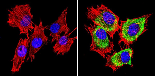 Immunocytochemistry/ Immunofluorescence - Anti-SREBP1 antibody (ab28481)
Immunocytochemical analysis of formalin-fixed C2C12 cell lines using immunofluorescence to label SREBP1 with ab28481 at a concentration of 1/100 in 3% BSA-PBS and incubated overnight in a humid environment at 4°C. Prior to labelling, cells were permeablised with 0.1% Triton X-100 in TBS for between 5 and 10 minutes, they were subsequently blocked with 3% BSA-PBS for 30 minutes at room temperature. The secondary used was a DyLight® conjugate (green) and was incubated at room temperature in the dark. The cells were counterstained with DAPI against DNA labelling the nucear compartments blue and a red fluorescent phalloidin stain against F-Actin. Magnification was 60X
The left image is a negative control in the absence of ab28481, the right image is in the prescence of ab28481, the secondary and counterstains. -
 Immunocytochemistry/ Immunofluorescence - Anti-SREBP1 antibody (ab28481)
Immunocytochemical analysis of formalin-fixed NIH 3T3 cell lines using immunofluorescence to label SREBP1 with ab28481 at a concentration of 1/100 in 3% BSA-PBS and incubated overnight in a humid environment at 4°C. Prior to labelling, cells were permeablised with 0.1% Triton X-100 in TBS for between 5 and 10 minutes, they were subsequently blocked with 3% BSA-PBS for 30 minutes at room temperature. The secondary used was a DyLight® conjugate (green) and was incubated at room temperature in the dark. The cells were counterstained with DAPI against DNA labelling the nucear compartments blue and a red fluorescent phalloidin stain against F-Actin. Magnification is 60X
The left image is a negative control in the absence of ab28481, the right image is in the prescence of ab28481, the secondary and counterstains.


