Anti-Semaphorin 7a antibody (ab23578)
Key features and details
- Rabbit polyclonal to Semaphorin 7a
- Suitable for: WB, ICC/IF, IP
- Reacts with: Mouse, Rat
- Isotype: IgG
Overview
-
Product name
Anti-Semaphorin 7a antibody
See all Semaphorin 7a primary antibodies -
Description
Rabbit polyclonal to Semaphorin 7a -
Host species
Rabbit -
Specificity
ab23578 detects Semaphorin 7a protein at 75 kDa by WB. Semaphorin 7a is a protein that is tethered to the cell membrane via a GPI-anchor and is also glycosylated, therefore it is not surprising that in WB we see some additional bands at ~50 and 100 kDa which are likely to be cleavage fragments and glycosylated protein respectively. Preincubation with the immunizing peptide ab30844 is successful in blocking antibody staining. -
Tested applications
Suitable for: WB, ICC/IF, IPmore details -
Species reactivity
Reacts with: Mouse, Rat
Predicted to work with: Cow
-
Immunogen
Synthetic peptide conjugated to KLH derived from within residues 1 - 100 of Mouse Semaphorin 7a.
Read Abcam's proprietary immunogen policy (Peptide available as ab30844.) -
General notes
Negative control: HEK cells
Properties
-
Form
Liquid -
Storage instructions
Shipped at 4°C. Store at +4°C short term (1-2 weeks). Upon delivery aliquot. Store at -20°C or -80°C. Avoid freeze / thaw cycle. -
Storage buffer
pH: 7.40
Preservative: 0.02% Sodium azide
Constituent: PBS
Batches of this product that have a concentration Concentration information loading...
Purity
Immunogen affinity purifiedClonality
PolyclonalIsotype
IgGResearch areas
Associated products
-
Compatible Secondaries
-
Isotype control
-
Recombinant Protein
Applications
Our Abpromise guarantee covers the use of ab23578 in the following tested applications.
The application notes include recommended starting dilutions; optimal dilutions/concentrations should be determined by the end user.
Application Abreviews Notes WB Use a concentration of 1 µg/ml. Detects a band of approximately 50,75,100 kDa (predicted molecular weight: 75 kDa). ICC/IF Use a concentration of 1 - 25 µg/ml. IP Use at an assay dependent concentration. Target
-
Function
Plays an important role in integrin-mediated signaling and functions both in regulating cell migration and immune responses. Promotes formation of focal adhesion complexes, activation of the protein kinase PTK2/FAK1 and subsequent phosphorylation of MAPK1 and MAPK3. Promotes production of proinflammatory cytokines by monocytes and macrophages. Plays an important role in modulating inflammation and T-cell-mediated immune responses. Promotes axon growth in the embryonic olfactory bulb. Promotes attachment, spreading and dendrite outgrowth in melanocytes. -
Tissue specificity
Detected in skin keratinocytes and on endothelial cells from skin blood vessels (at protein level). Expressed in fibroblasts, keratinocytes, melanocytes, placenta, testis, ovary, spleen, brain, spinal chord, lung, heart, adrenal gland, lymph nodes, thymus, intestine and kidney. -
Sequence similarities
Belongs to the semaphorin family.
Contains 1 Ig-like C2-type (immunoglobulin-like) domain.
Contains 1 PSI domain.
Contains 1 Sema domain. -
Cellular localization
Cell membrane. Detected in a punctate pattern on the cell membrane of basal and supra-basal skin keratinocytes. - Information by UniProt
-
Database links
- Entrez Gene: 20361 Mouse
- Entrez Gene: 315711 Rat
- SwissProt: Q9QUR8 Mouse
- Unigene: 335187 Mouse
-
Alternative names
- CD 108 antibody
- CD108 antibody
- CD108 antigen antibody
see all
Images
-
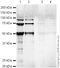
All lanes : Anti-Semaphorin 7a antibody (ab23578) at 1 µg/ml
Lane 1 : Brain (Mouse) Tissue Lysate (ab27253)
Lane 2 :Mouse brain tissue lysate - total protein (0 days) (ab7188)
Lane 3 : Brain (Mouse) Tissue Lysate (ab27253) with Mouse Semaphorin 7a peptide (ab30844) at 1 µg/ml
Lane 4 :Mouse brain tissue lysate - total protein (0 days) (ab7188) with Mouse Semaphorin 7a peptide (ab30844) at 1 µg/ml
Lysates/proteins at 20 µg per lane.
Secondary
All lanes : Rabbit IgG secondary antibody (ab28446) at 1/10000 dilution
Performed under reducing conditions.
Predicted band size: 75 kDa
Observed band size: 75 kDa
Additional bands at: 100 kDa (possible glycosylated form), 50 kDa (possible cleavage fragment)ab23578 detects Semaphorin 7a protein at 75kDa by WB. Semaphorin 7a is a protein that is tethered to the cell membrane via a GPI-anchor and is also glycosylated, therefore it is not surprising that we see some additional bands at ~50 and 100 kDa which are likely to be cleavage fragments and glycosylated protein respectively.
-
ICC/IF image of ab23578 stained PC12 cells. The cells were 4% formaldehyde fixed (10 min) and then incubated in 1%BSA / 10% normal goat serum / 0.3M glycine in 0.1% PBS-Tween for 1h to permeabilise the cells and block non-specific protein-protein interactions. The cells were then incubated with the antibody (ab23578, 1µg/ml) overnight at +4°C. The secondary antibody (green) was Alexa Fluor® 488 goat anti-rabbit IgG (H+L) used at a 1/1000 dilution for 1h. Alexa Fluor® 594 WGA was used to label plasma membranes (red) at a 1/200 dilution for 1h. DAPI was used to stain the cell nuclei (blue).
-
 Immunocytochemistry/ Immunofluorescence - Anti-Semaphorin 7a antibody (ab23578)This image is courtesy of Randal Moldrich, CNRS UMR7637, ESPCI, France
ab23578 detecting Semaphorin 7a in developing mouse neurons, differentiated from neural precursor cells cultured from embryonic day 13 neocortex; neurons were cultured on poly-D-lysine in the presence of Neurobasal Medium containing B27 supplement. Immunocytochemistry: all steps were performed in PBS. Cultures were fixed in 4% PFA for 15min, permeabilised with 0.1% Triton X-100 for 10min and blocked with 5% BSA, 0.1% Triton X-100 for 45min. ab23578 was incubated at 5µg/ml (12h in 5% BSA, 0.1% Triton X-100) at 4°C. Goat anti-rabbit AlexaFluor 488 was used as secondary antibody at 1/400 for 1hr at RT (in 5% BSA, 0.1% Triton X-100). Treated cultures were mounted on glass coverslips with Mowiol. ab23578 immunostaining (green colour) was neuronal and for the most part at the membrane with some cytosolic staining observed. No nuclear staining was observed, confirmed with a nuclear counterstain (To-pro-3; data not shown).
-
Semaphorin 7a was immunoprecipitated using 0.5mg Mouse Brain whole tissue lysate, 5µg of Rabbit polyclonal to Semaphorin 7a and 50µl of protein G magnetic beads (+). No antibody was added to the control (-).
The antibody was incubated under agitation with Protein G beads for 10min, Mouse Brain whole tissue lysate lysate diluted in RIPA buffer was added to each sample and incubated for a further 10min under agitation.
Proteins were eluted by addition of 40µl SDS loading buffer and incubated for 10min at 70oC; 10µl of each sample was separated on a SDS PAGE gel, transferred to a nitrocellulose membrane, blocked with 5% BSA and probed with ab23578.
Secondary: Mouse monoclonal [SB62a] Secondary Antibody to Rabbit IgG light chain (HRP) (ab99697).
Band: 75kDa: Semaphorin 7a.
Protocols
References (17)
ab23578 has been referenced in 17 publications.
- Köhler D et al. Red blood cell-derived semaphorin 7A promotes thrombo-inflammation in myocardial ischemia-reperfusion injury through platelet GPIb. Nat Commun 11:1315 (2020). PubMed: 32161256
- Hu S et al. Semaphorin 7A Promotes VEGFA/VEGFR2-Mediated Angiogenesis and Intraplaque Neovascularization in ApoE-/- Mice. Front Physiol 9:1718 (2018). PubMed: 30555351
- Chen X et al. Anti-Semaphorin-7A single chain antibody demonstrates beneficial effects on pulmonary inflammation during acute lung injury. Exp Ther Med 15:2356-2364 (2018). PubMed: 29456642
- Chabrat A et al. Transcriptional repression of Plxnc1 by Lmx1a and Lmx1b directs topographic dopaminergic circuit formation. Nat Commun 8:933 (2017). PubMed: 29038581
- Jongbloets BC et al. Stage-specific functions of Semaphorin7A during adult hippocampal neurogenesis rely on distinct receptors. Nat Commun 8:14666 (2017). WB ; Mouse . PubMed: 28281529
Images
-
All lanes : Anti-Semaphorin 7a antibody (ab23578) at 1 µg/ml
Lane 1 : Brain (Mouse) Tissue Lysate (ab27253)
Lane 2 :Mouse brain tissue lysate - total protein (0 days) (ab7188)
Lane 3 : Brain (Mouse) Tissue Lysate (ab27253) with Mouse Semaphorin 7a peptide (ab30844) at 1 µg/ml
Lane 4 :Mouse brain tissue lysate - total protein (0 days) (ab7188) with Mouse Semaphorin 7a peptide (ab30844) at 1 µg/ml
Lysates/proteins at 20 µg per lane.
Secondary
All lanes : Rabbit IgG secondary antibody (ab28446) at 1/10000 dilution
Performed under reducing conditions.
Predicted band size: 75 kDa
Observed band size: 75 kDa
Additional bands at: 100 kDa (possible glycosylated form), 50 kDa (possible cleavage fragment)ab23578 detects Semaphorin 7a protein at 75kDa by WB. Semaphorin 7a is a protein that is tethered to the cell membrane via a GPI-anchor and is also glycosylated, therefore it is not surprising that we see some additional bands at ~50 and 100 kDa which are likely to be cleavage fragments and glycosylated protein respectively.
-
ICC/IF image of ab23578 stained PC12 cells. The cells were 4% formaldehyde fixed (10 min) and then incubated in 1%BSA / 10% normal goat serum / 0.3M glycine in 0.1% PBS-Tween for 1h to permeabilise the cells and block non-specific protein-protein interactions. The cells were then incubated with the antibody (ab23578, 1µg/ml) overnight at +4°C. The secondary antibody (green) was Alexa Fluor® 488 goat anti-rabbit IgG (H+L) used at a 1/1000 dilution for 1h. Alexa Fluor® 594 WGA was used to label plasma membranes (red) at a 1/200 dilution for 1h. DAPI was used to stain the cell nuclei (blue).
-
 Immunocytochemistry/ Immunofluorescence - Anti-Semaphorin 7a antibody (ab23578) This image is courtesy of Randal Moldrich, CNRS UMR7637, ESPCI, France
ab23578 detecting Semaphorin 7a in developing mouse neurons, differentiated from neural precursor cells cultured from embryonic day 13 neocortex; neurons were cultured on poly-D-lysine in the presence of Neurobasal Medium containing B27 supplement. Immunocytochemistry: all steps were performed in PBS. Cultures were fixed in 4% PFA for 15min, permeabilised with 0.1% Triton X-100 for 10min and blocked with 5% BSA, 0.1% Triton X-100 for 45min. ab23578 was incubated at 5µg/ml (12h in 5% BSA, 0.1% Triton X-100) at 4°C. Goat anti-rabbit AlexaFluor 488 was used as secondary antibody at 1/400 for 1hr at RT (in 5% BSA, 0.1% Triton X-100). Treated cultures were mounted on glass coverslips with Mowiol. ab23578 immunostaining (green colour) was neuronal and for the most part at the membrane with some cytosolic staining observed. No nuclear staining was observed, confirmed with a nuclear counterstain (To-pro-3; data not shown).
-
Semaphorin 7a was immunoprecipitated using 0.5mg Mouse Brain whole tissue lysate, 5µg of Rabbit polyclonal to Semaphorin 7a and 50µl of protein G magnetic beads (+). No antibody was added to the control (-).
The antibody was incubated under agitation with Protein G beads for 10min, Mouse Brain whole tissue lysate lysate diluted in RIPA buffer was added to each sample and incubated for a further 10min under agitation.
Proteins were eluted by addition of 40µl SDS loading buffer and incubated for 10min at 70oC; 10µl of each sample was separated on a SDS PAGE gel, transferred to a nitrocellulose membrane, blocked with 5% BSA and probed with ab23578.
Secondary: Mouse monoclonal [SB62a] Secondary Antibody to Rabbit IgG light chain (HRP) (ab99697).
Band: 75kDa: Semaphorin 7a.


