Anti-RALBP1 antibody (ab33446)
")
Key features and details
- Rabbit polyclonal to RALBP1
- Suitable for: ICC/IF, WB
- Reacts with: Human
- Isotype: IgG
Overview
-
Product name
Anti-RALBP1 antibody
See all RALBP1 primary antibodies -
Description
Rabbit polyclonal to RALBP1 -
Host species
Rabbit -
Tested Applications & Species
See all applications and species dataApplication Species ICC/IF HumanWB Human
-
Immunogen
Synthetic peptide corresponding to Human RALBP1 aa 600 to the C-terminus (C terminal) conjugated to keyhole limpet haemocyanin.
(Peptide available asab33445) -
Positive control
- WB: Jurkat, HEK293, Hap1, HeLa and A431 cell lysates. ICC/IF: HEK293 cells.
Properties
-
Form
Liquid -
Storage instructions
Shipped at 4°C. Store at +4°C short term (1-2 weeks). Upon delivery aliquot. Store at -20°C or -80°C. Avoid freeze / thaw cycle. -
Storage buffer
pH: 7.40
Preservative: 0.02% Sodium azide
Constituent: PBS
Batches of this product that have a concentration Concentration information loading...
Purity
Immunogen affinity purifiedClonality
PolyclonalIsotype
IgGResearch areas
Associated products
-
Compatible Secondaries
-
Isotype control
-
KO cell lines
-
KO cell lysates
Applications
The Abpromise guarantee
Our Abpromise guarantee covers the use of ab33446 in the following tested applications.
The application notes include recommended starting dilutions; optimal dilutions/concentrations should be determined by the end user.
GuaranteedTested applications are guaranteed to work and covered by our Abpromise guarantee.
PredictedPredicted to work for this combination of applications and species but not guaranteed.
IncompatibleDoes not work for this combination of applications and species.
Application Species ICC/IF HumanWB HumanAll applications RatApplication Abreviews Notes ICC/IF Use a concentration of 1 µg/ml.WB (1) Use a concentration of 1 µg/ml. Detects a band of approximately 90 kDa (predicted molecular weight: 76 kDa).Notes ICC/IF
Use a concentration of 1 µg/ml.WB
Use a concentration of 1 µg/ml. Detects a band of approximately 90 kDa (predicted molecular weight: 76 kDa).Target
-
Function
Can activate specifically hydrolysis of GTP bound to RAC1 and CDC42, but not RALA. Mediates ATP-dependent transport of S-(2,4-dinitrophenyl)-glutathione (DNP-SG) and doxorubicin (DOX) and is the major ATP-dependent transporter of glutathione conjugates of electrophiles (GS-E) and DOX in erythrocytes. Can catalyze transport of glutathione conjugates and xenobiotics, and may contribute to the multidrug resistance phenomenon. Serves as a scaffold protein that brings together proteins forming an endocytotic complex during interphase and also with CDK1 to switch off endocytosis, One of its substrates would be EPN1/Epsin. -
Tissue specificity
Expressed ubiquitously but at low levels. Shows a strong expression in the erythrocytes. -
Sequence similarities
Contains 1 Rho-GAP domain. -
Cellular localization
Membrane. - Information by UniProt
-
Database links
- Entrez Gene: 10928 Human
- Entrez Gene: 84014 Rat
- Omim: 605801 Human
- SwissProt: Q15311 Human
- SwissProt: Q62796 Rat
- Unigene: 528993 Human
- Unigene: 7107 Rat
-
Alternative names
- RLIP1 antibody
- 76 kDa Ral-interacting protein antibody
- 76-kDa Ral-interacting protein antibody
see all
Images
-
Western blot - Anti-RALBP1 antibody (ab33446)All lanes : Anti-RALBP1 antibody (ab33446) at 1/1000 dilution
Lane 1 : Wild-type HeLa cell lysate
Lane 2 : RALBP1 knockout HeLa cell lysate
Lane 3 : HAP1 cell lysate
Lane 4 : Jurkat cell lysate
Lysates/proteins at 20 µg per lane.
Performed under reducing conditions.
Predicted band size: 76 kDaLanes 1-4: Merged signal (red and green). Green - ab33446 observed at 90 kDa. Red - loading control ab8245 observed at 37 kDa.
ab33446 Anti-RALBP1 antibody was shown to specifically react with PDE10A in wild-type HeLa cells. Loss of signal was observed when knockout cell line ab265404 (knockout cell lysate ab258167) was used. Wild-type and PDE10A knockout samples were subjected to SDS-PAGE. ab33446 and Anti-GAPDH antibody [6C5] - Loading Control (ab8245) were incubated overnight at 4°C at 1 in 1000 dilution and 1 in 20000 dilution respectively. Blots were developed with Goat anti-Rabbit IgG H&L (IRDye® 800CW) preadsorbed (ab216773) and Goat anti-Mouse IgG H&L (IRDye® 680RD) preadsorbed (ab216776) secondary antibodies at 1 in 20000 dilution for 1 hour at room temperature before imaging.
-
 Western blot - Anti-RALBP1 antibody (ab33446)All lanes : Anti-RALBP1 antibody (ab33446) at 1 µg/ml
Lane 1 : Jurkat (Human T cell lymphoblast-like cell line) Whole Cell Lysate
Lane 2 : HEK293 (Human embryonic kidney cell line) Whole Cell Lysate
Lysates/proteins at 10 µg per lane.
Secondary
All lanes : IRDye 680 Conjugated Goat Anti-Rabbit IgG (H+L) at 1/10000 dilution
Performed under reducing conditions.
Predicted band size: 76 kDa
Observed band size: 90 kDa why is the actual band size different from the predicted?
The band seen at 90 kDa is consistent with the banding pattern observed for other commercially available antibodies to RIP1. -
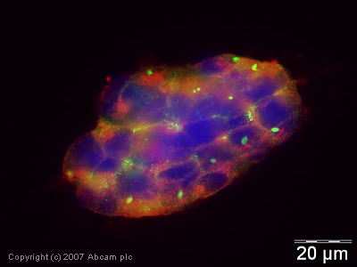 Immunocytochemistry/ Immunofluorescence - Anti-RALBP1 antibody (ab33446)ICC/IF image of ab33446 stained human HEK 293 cells. The cells were methanol fixed (5 min), permabilised in TBS-T (20 min) and incubated with the antibody (ab33446, 1µg/ml) for 1h at room temperature. 1%BSA / 10% normal goat serum / 0.3M glycine was used to quench autofluorescence and block non-specific protein-protein interactions. The secondary antibody (green) was Alexa Fluor® 488 goat anti-rabbit IgG (H+L) used at a 1/1000 dilution for 1h. Alexa Fluor® 594 WGA was used to label plasma membranes (red). DAPI was used to stain the cell nuclei (blue).
Protocols
Datasheets and documents
References (1)
ab33446 has been referenced in 1 publication.
- Johansson J et al. RAL GTPases Drive Intestinal Stem Cell Function and Regeneration through Internalization of WNT Signalosomes. Cell Stem Cell 24:592-607.e7 (2019). PubMed: 30853556
Images
-
Western blot - Anti-RALBP1 antibody (ab33446)All lanes : Anti-RALBP1 antibody (ab33446) at 1/1000 dilution
Lane 1 : Wild-type HeLa cell lysate
Lane 2 : RALBP1 knockout HeLa cell lysate
Lane 3 : HAP1 cell lysate
Lane 4 : Jurkat cell lysate
Lysates/proteins at 20 µg per lane.
Performed under reducing conditions.
Predicted band size: 76 kDaLanes 1-4: Merged signal (red and green). Green - ab33446 observed at 90 kDa. Red - loading control ab8245 observed at 37 kDa.
ab33446 Anti-RALBP1 antibody was shown to specifically react with PDE10A in wild-type HeLa cells. Loss of signal was observed when knockout cell line ab265404 (knockout cell lysate ab258167) was used. Wild-type and PDE10A knockout samples were subjected to SDS-PAGE. ab33446 and Anti-GAPDH antibody [6C5] - Loading Control (ab8245) were incubated overnight at 4°C at 1 in 1000 dilution and 1 in 20000 dilution respectively. Blots were developed with Goat anti-Rabbit IgG H&L (IRDye® 800CW) preadsorbed (ab216773) and Goat anti-Mouse IgG H&L (IRDye® 680RD) preadsorbed (ab216776) secondary antibodies at 1 in 20000 dilution for 1 hour at room temperature before imaging.
-
Western blot - Anti-RALBP1 antibody (ab33446)All lanes : Anti-RALBP1 antibody (ab33446) at 1 µg/ml
Lane 1 : Jurkat (Human T cell lymphoblast-like cell line) Whole Cell Lysate
Lane 2 : HEK293 (Human embryonic kidney cell line) Whole Cell Lysate
Lysates/proteins at 10 µg per lane.
Secondary
All lanes : IRDye 680 Conjugated Goat Anti-Rabbit IgG (H+L) at 1/10000 dilution
Performed under reducing conditions.
Predicted band size: 76 kDa
Observed band size: 90 kDa why is the actual band size different from the predicted?
The band seen at 90 kDa is consistent with the banding pattern observed for other commercially available antibodies to RIP1. -
Immunocytochemistry/ Immunofluorescence - Anti-RALBP1 antibody (ab33446)ICC/IF image of ab33446 stained human HEK 293 cells. The cells were methanol fixed (5 min), permabilised in TBS-T (20 min) and incubated with the antibody (ab33446, 1µg/ml) for 1h at room temperature. 1%BSA / 10% normal goat serum / 0.3M glycine was used to quench autofluorescence and block non-specific protein-protein interactions. The secondary antibody (green) was Alexa Fluor® 488 goat anti-rabbit IgG (H+L) used at a 1/1000 dilution for 1h. Alexa Fluor® 594 WGA was used to label plasma membranes (red). DAPI was used to stain the cell nuclei (blue).


