Anti-Rab3A antibody (ab3335)
Key features and details
- Rabbit polyclonal to Rab3A
- Suitable for: WB, ICC/IF
- Reacts with: Mouse, Rat, Human
- Isotype: IgG
Overview
-
Product name
Anti-Rab3A antibody
See all Rab3A primary antibodies -
Description
Rabbit polyclonal to Rab3A -
Host species
Rabbit -
Tested applications
Suitable for: WB, ICC/IFmore details -
Species reactivity
Reacts with: Mouse, Rat, Human -
Immunogen
Synthetic peptide corresponding to Human Rab3A aa 1-18.
Sequence:MASATDSRYGQKESSDQN
(Peptide available asab4951)
Properties
-
Form
Liquid -
Storage instructions
Shipped at 4°C. Store at +4°C short term (1-2 weeks). Upon delivery aliquot. Store at -20°C or -80°C. Avoid freeze / thaw cycle. -
Storage buffer
Preservative: 0.05% Sodium azide
Constituent: 0.1% BSA -
 Concentration information loading...
 -
Purity
Protein A purified -
Primary antibody notes
Rab proteins are low-molecular-weight GTP-binding proteins that form the largest branch of the Ras superfamily of GTPases. Located on the cytoplasmic face of organelles and vesicles, rab proteins are involved in intracellular membrane fusion reactions. Three membrane proteins, synaptosomal associated protein of 25 kDa (SNAP-25), synaptobrevin and syntaxin, form the core of a ubiquitous membrane fusion machine that interacts with the soluble proteins N-ethylmaleimide-sensitive factor (NSF) and a-SNAP. Rab proteins, in co-ordination with the core fusion machinery and Munc-18, help to mediate vesicle docking and fusion. There exist over 40 rab proteins that have been described in mammals and the best studied is rab 3A. Rab 3A is found to be abundant in presynaptic nerve terminals and is also found to be crucial in acrosomal exocytosis in human spermatozoa. Abnormal accumulation of rab 3A in the cytoplasm of Purkinje cells has been reported in the prion protein related Creutzfeldt-Jakob disease. -
Clonality
Polyclonal -
Isotype
IgG -
Research areas
Images
-
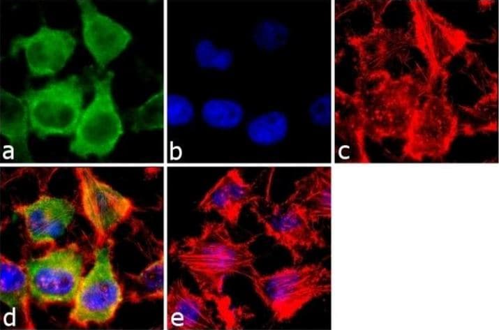
Immunofluorescence analysis of RAB3A was done on 70% confluent log phase HeLa (Human epithelial adenocarcinoma cell line)
cells. The cells were fixed with 4% paraformaldehyde for 10 minutes, permeabilized with 0.1% Triton™ X-100 for 10 minutes, and blocked with 1% BSA for 1 hour at room temperature. The cells were labeled with ab3335 at 2 µg/mL in 0.1% BSA and incubated for 3 hours at room temperature and then labeled with Goat anti-Rabbit IgG (H+L) Superclonal™ Secondary Antibody, Alexa Fluor® 488 conjugate at 1/2000 dilution for 45 minutes at room temperature (Panel a: green). Nuclei (Panel b: blue) were stained with SlowFade® Gold Antifade Mountant with DAPI. F-actin (Panel c: red) was stained with Alexa Fluor® 555 Rhodamine Phalloidin at 1/300 dilution. Panel d is a merged image showing Cytoplasmic localization. Panel e is a no primary antibody control. The images were captured at 60X magnification. -
All lanes : Anti-Rab3A antibody (ab3335) at 2 µg/ml
Lane 1 : Mouse brain tissue lysate
Lane 2 : Mouse kidney tissue lysate
Lane 3 : Mouse liver tissue lysate
Lane 4 : Rat brain tissue lysate
Lane 5 : Rat kidney tissue lysate
Lane 6 : Rat liver tissue lysate
Lysates/proteins at 30 µg per lane.
Secondary
All lanes : Rabbit anti-Goat IgG (H+L) Superclonal™ Recombinant Secondary Antibody, HRP at 1/4000 dilutionA 27 kDa band corresponding to RAB3A was observed along with two uncharacterized band (*) at ~40 kD and 60 kDa across tissues tested.


