Anti-PYK2 (phospho Y579 + Y580) antibody (ab4807)
Key features and details
- Rabbit polyclonal to PYK2 (phospho Y579 + Y580)
- Suitable for: WB
- Reacts with: Chicken
- Isotype: IgG
Overview
-
Product name
Anti-PYK2 (phospho Y579 + Y580) antibody
See all PYK2 primary antibodies -
Description
Rabbit polyclonal to PYK2 (phospho Y579 + Y580) -
Host species
Rabbit -
Specificity
Some cross-reactivity still may be experienced in cases where Focal Adhesion Kinase is overexpressed relative to PYK 2 within the same cell system. -
Tested Applications & Species
See all applications and species dataApplication Species WB Chicken -
Immunogen
Synthetic peptide (Human) derived from the region of human Pyk 2 that contains tyrosines 579 and 580. The sequence is conserved in human and rat.
-
Positive control
- Vandate treated CEF cells expressing PYK 2 and plated on fibronectin.
Properties
-
Form
Liquid -
Storage instructions
Shipped at 4°C. Upon delivery aliquot and store at -20°C or -80°C. Avoid repeated freeze / thaw cycles. -
Storage buffer
pH: 7.3
Constituents: PBS, 50% Glycerol (glycerin, glycerine), 1% BSA -
 Concentration information loading...
 -
Purity
Immunogen affinity purified -
Purification notes
Purified from rabbit serum by sequential epitope-specific chromatography. The antibody has been negatively preadsorbed using (i) a non-phosphopeptide corresponding to the site of phosphorylation to remove antibody that is reactive with non-phosphorylated PYK 2, (ii) a generic tyrosine phosphorylated peptide to remove antibody that is reactive with phosphotyrosine, irrespective of the sequence, and (iii) a phosphopeptide derived from the corresponding region of Focal Adhesion Kinase (a PYK 2-related protein) to remove antibody that is reactive with phosphorylated Focal Adhesion Kinase protein. The final product is generated by affinity chromatography using a PYK 2-derived peptide that is phosphorylated at tyrosines 579 and 580. -
Clonality
Polyclonal -
Isotype
IgG -
Research areas
Images
-
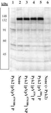
Peptide Competition: Cell extracts prepared from chick embryo fibroblasts treated with vanadate were plated on fibronectin with (lanes 1-5) or without (lane 6) transfection of a PYK 2 expression vector and resolved by SDS PAGE on a 10% Tris-glycine gel. The proteins were then transferred to nitrocellulose. Membranes were incubated with 0.50
µ g/mL ab4807, following prior incubation in the presence of the phosphopeptide immunogen (1), the absence of the phosphopeptide immunogen (2, 6), the non phosphopeptide corresponding to the PYK 2 phosphopeptide (3), the phosphopeptide corresponding to PYK 2 [pY579] (4), and the phosphopeptide corresponding to PYK 2 [pY580] (5). After washing, membranes were incubated with goat F(ab’)2 anti-rabbit IgG alkaline phosphatase and signals were detected using the Tropix WesternStar method. The data show that only the dual-phosphopeptide corresponding to this site blocks the antibody signal, not the corresponding mono phosphopeptides, t


