Anti-PMP70 antibody (ab3421)
")
Key features and details
- Rabbit polyclonal to PMP70
- Suitable for: ICC/IF, IHC-P, Flow Cyt, WB
- Reacts with: Mouse, Rat, Human
- Isotype: IgG
Overview
-
Product name
Anti-PMP70 antibody
See all PMP70 primary antibodies -
Description
Rabbit polyclonal to PMP70 -
Host species
Rabbit -
Tested Applications & Species
See all applications and species dataApplication Species Flow Cyt MouseHumanICC/IF MouseHumanIHC-P HumanWB MouseRat
-
Immunogen
Synthetic peptide corresponding to Rat PMP70 aa 644-659.
Sequence:NYEFKKITEDTVEFGS
(Peptide available asab4965) -
Positive control
- WB: Rat kidney and liver tissue lysate, A431, U-2 OS, HepG2 whole cell lysates. ICC: A431 KO, A431, NIH/3T3, HMVEC, NS-1, P19, A549 whole cells. IHC-P: Human duodenum tissue.
-
General notes
The Life Science industry has been in the grips of a reproducibility crisis for a number of years. Abcam is leading the way in addressing the problem with our range of recombinant monoclonal antibodies and knockout edited cell lines for gold-standard validation.
One factor contributing to the crisis is the use of antibodies that are not suitable. This can lead to misleading results and the use of incorrect data informing project assumptions and direction. To help address this challenge, we have introduced an application and species grid on our primary antibody datasheets to make it easy to simplify identification of the right antibody for your needs.
Learn more here.


Properties
-
Form
Liquid -
Storage instructions
Shipped at 4°C. Store at +4°C short term (1-2 weeks). Upon delivery aliquot. Store at -20°C or -80°C. Avoid freeze / thaw cycle. -
Storage buffer
Constituents: 0.1% BSA, 99% PBS -
 Concentration information loading...
 -
Purity
Immunogen affinity purified -
Clonality
Polyclonal -
Isotype
IgG -
Research areas
Images
-
Western blot - Anti-PMP70 antibody (ab3421)All lanes : Anti-PMP70 antibody (ab3421) at 1/500 dilution
Lane 1 : Rat kidney tissue lysate at 20 µg
Lane 2 : Rat liver tissue lysate at 20 µg
Lane 3 : Mouse lung tissue lysate at 20 µg
Lane 4 : A431 (Human epidermoid carcinoma cell line) whole cell lysate at 10 µg
Lane 5 : U-2 OS (Human bone osteosarcoma epithelial cell line) whole cell lysate at 10 µg
-
 Immunocytochemistry/ Immunofluorescence - Anti-PMP70 antibody (ab3421)
Knockdown of PMP70 was achieved by transfecting A431 (Human epidermoid carcinoma cell line) cells with PMP70 specific siRNA. Immunofluorescence analysis was performed on A-431 cells (untransfected, panel a,d), transfected with non-specific scrambled siRNA (panels b,e) and transfected with PMP70 specific siRNA (panel c,f). Cells were fixed, permeabilized, and labelled with PMP70 Rabbit Polyclonal Antibody (5 µg/ml), followed by Goat anti-Rabbit IgG (H+L) Superclonal™ Recombinant Secondary Antibody, Alexa Fluor® 488 (1:2000). Nuclei (blue) were stained using ProLong™ Diamond Antifade Mountant with DAPI, and Rhodamine Phalloidin (1:300) was used for cytoskeletal F-actin (red) staining. Reduction of specific signal was observed upon siRNA mediated knockdown (panel c,f) confirming specificity of the antibody to PMP70 (green). The images were captured at 60X magnification.
-
 Flow Cytometry - Anti-PMP70 antibody (ab3421)
Flow cytometry analysis of PMP70 in HEK-293T (Human epithelial cell line from embryonic kidney transformed with large T antigen) cells (green) compared to an isotype control (blue). Cells were harvested, adjusted to a concentration of 1-5x10⁶ cells/ml, fixed with 2% paraformaldehyde and washed with PBS. Cells were blocked with a 2% solution of BSA-PBS for 30 min at room temperature and incubated with ab3421 at a dilution of 0.5 ug/test for 60 min at room temperature. Cells were then incubated for 40 min at room temperature in the dark using a Dylight 488-conjugated goat anti-mouse IgG (H+L) secondary antibody and re-suspended in PBS for FACS analysis.
-
 Immunohistochemistry (Formalin/PFA-fixed paraffin-embedded sections) - Anti-PMP70 antibody (ab3421)ab3421 (2µg/ml) staining PMP70 in human duodenum using an automated system (DAKO Autostainer Plus). Using this protocol there is apical cytoplasmic staining and staining of the endoplasmic reticulum in the epithelium.
Sections were rehydrated and antigen retrieved with the Dako 3 in 1 AR buffer EDTA pH 9.0 in a DAKO PT Link. Slides were peroxidase blocked in 3% H2O2 in methanol for 10 mins. They were then blocked with Dako Protein block for 10 minutes (containing casein 0.25% in PBS) then incubated with primary antibody for 20 min and detected with Dako Envision Flex amplification kit for 30 minutes. Colorimetric detection was completed with Diaminobenzidine for 5 minutes. Slides were counterstained with Haematoxylin and coverslipped under DePeX. Please note that, for manual staining, optimization of primary antibody concentration and incubation time is recommended. Signal amplification may be required.
-
 Western blot - Anti-PMP70 antibody (ab3421)All lanes : Anti-PMP70 antibody (ab3421) at 1/1000 dilution
Lane 1 : Mouse lung tissue lysate
Lane 2 : Mouse liver tissue lysate
Lane 3 : HepG2 (Human liver hepatocellular carcinoma cell line) whole cell lysate
Lysates/proteins at 25 µg per lane.
Observed band size: 70 kDa why is the actual band size different from the predicted?
-
 Immunocytochemistry/ Immunofluorescence - Anti-PMP70 antibody (ab3421)
Immunofluorescent analysis of PMP70 (green) in NIH/3T3 (Mouse embryo fibroblast cell line) ells. The cells were permeabilized with 0.1% Triton X-100 in TBS for 15 minutes, and blocked with 3% Blocker BSA in PBS for 15 minutes at room temperature. Cells were stained with ab3421 at 10 µg/mL in blocking buffer for at least 1 hour at room temperature, and then incubated with a Goat anti-Rabbit IgG Superclonal secondary antibody, Alexa Fluor 488 conjugate at a dilution of 1:1000 for 30 minutes at room temperature (green). Nuclei (blue) were stained with Hoechst 33342 dye. Images were taken on a Thermo Scientific ToxInsight Instrument at 20X magnification.
-
 Flow Cytometry - Anti-PMP70 antibody (ab3421)
Flow cytometry analysis of PMP70 in NIH/3T3 (Mouse embryo fibroblast cell line) whole cells (green) compared to an isotype control (blue). Cells were harvested, adjusted to a concentration of 1-5x10⁶ cells/ml, fixed with 2% paraformaldehyde and washed with PBS. Cells were blocked with a 2% solution of BSA-PBS for 30 min at room temperature and incubated with ab3421 at a dilution of 0.5 ug/test for 60 min at room temperature. Cells were then incubated for 40 min at room temperature in the dark using a Dylight 488-conjugated goat anti-mouse IgG (H+L) secondary antibody and re-suspended in PBS for FACS analysis.
-
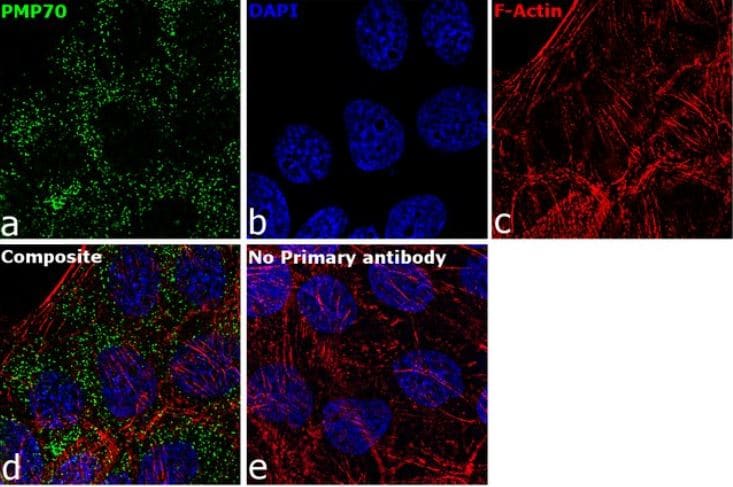 Immunocytochemistry/ Immunofluorescence - Anti-PMP70 antibody (ab3421)
Immunofluorescence analysis of PMP70 was performed using 70% confluent log phase A431 (Human epidermoid carcinoma cell line) cells. The cells were fixed with 4% paraformaldehyde for 10 minutes, permeabilized with 0.1% Triton™ X-100 for 15 minutes, and blocked with 2% BSA for 1 hour at room temperature. The cells were labeled with ab3421 at 5 µg/ml in 0.1% BSA, incubated at 4 degree celsius overnight and then labeled with Goat anti-Rabbit IgG (H+L), Superclonal™ Recombinant Secondary Antibody, Alexa Fluor 488 at a dilution of 1:2000 for 45 minutes at room temperature (Panel a: green). Nuclei (Panel b: blue) were stained with ProLong™ Diamond Antifade Mountant with DAPI. F-actin (Panel c: red) was stained with Rhodamine Phalloidin. Panel d represents the merged image showing cytoplasmic (peroxisomal pattern) localization. Panel e represents control cells with no primary antibody to assess background. The images were captured at 60X magnification.
-
 Flow Cytometry - Anti-PMP70 antibody (ab3421)
Flow cytometry analysis of PMP70 in HepG2 (Human liver hepatocellular carcinoma cell line) cells (green) compared to an isotype control (blue). Cells were harvested, adjusted to a concentration of 1-5x10⁶ cells/ml, fixed with 2% paraformaldehyde and washed with PBS. Cells were blocked with a 2% solution of BSA-PBS for 30 min at room temperature and incubated with ab3421 at a dilution of 0.5 ug/test for 60 min at room temperature. Cells were then incubated for 40 min at room temperature in the dark using a Dylight 488-conjugated goat anti-mouse IgG (H+L) secondary antibody and re-suspended in PBS for FACS analysis.
-
 Immunocytochemistry/ Immunofluorescence - Anti-PMP70 antibody (ab3421)
ICC/IF image of ab3421 stained human HeLa (Human epithelial adenocarcinoma cell line) cells. The cells were 4% PFA fixed (10 min) and then incubated in 1% BSA / 10% normal goat serum / 0.3M glycine in 0.1% PBS-Tween for 1h to permeabilise the cells and block non-specific protein-protein interactions. The cells were then incubated with the antibody (ab3421, 1 µg/ml) overnight at +4°C. The secondary antibody (green) was Alexa Fluor® 488 goat anti-rabbit IgG (H+L) used at a 1/1000 dilution for 1h. Alexa Fluor® 594 WGA was used to label plasma membranes (red) at a 1/200 dilution for 1h. DAPI was used to stain the cell nuclei (blue). This antibody also gave a positive IF result in Hek293, HepG2 and MCF7 cells.
-
 Immunocytochemistry/ Immunofluorescence - Anti-PMP70 antibody (ab3421)
Immunofluorescence analysis of PMP70 using ab3421 shows staining in HMVEC (Human microvascular endothelial cell line) cells.
-
 Immunocytochemistry/ Immunofluorescence - Anti-PMP70 antibody (ab3421)
Immunofluorescence analysis of PMP70 using ab3421 shows staining in NS-1 (Mouse myeloma cell line) cells.
-
 Immunocytochemistry/ Immunofluorescence - Anti-PMP70 antibody (ab3421)
Immunofluorescence analysis of PMP70 using ab3421 shows staining in P19 (Mouse embryonal carcinoma cell line) cells.
-
 Immunocytochemistry/ Immunofluorescence - Anti-PMP70 antibody (ab3421)
Immunofluorescence analysis of PMP70 using ab3421 shows staining in A549 (Human lung carcinoma cell line) cells.


