Anti-PKM2 antibody (ab85555)
Key features and details
- Rabbit polyclonal to PKM2
- Suitable for: WB, ICC/IF
- Reacts with: Human
- Isotype: IgG
Overview
-
Product name
Anti-PKM2 antibody
See all PKM2 primary antibodies -
Description
Rabbit polyclonal to PKM2 -
Host species
Rabbit -
Tested Applications & Species
See all applications and species dataApplication Species ICC/IF HumanWB Human -
Immunogen
Synthetic peptide corresponding to Human PKM2 aa 1-100 conjugated to keyhole limpet haemocyanin.
(Peptide available asab94378) -
Positive control
- This antibody gave a positive signal in the following whole cell lysates: HeLa; MEL-1; HepG2; MCF7; Caco 2; SHSY-5Y; Raji
Properties
-
Form
Liquid -
Storage instructions
Shipped at 4°C. Store at +4°C short term (1-2 weeks). Upon delivery aliquot. Store at -20°C or -80°C. Avoid freeze / thaw cycle. -
Storage buffer
pH: 7.40
Preservative: 0.02% Sodium azide
Constituent: PBS
Batches of this product that have a concentration Concentration information loading...
Purity
Immunogen affinity purifiedClonality
PolyclonalIsotype
IgGResearch areas
Associated products
-
Compatible Secondaries
-
Isotype control
-
Positive Controls
-
Recombinant Protein
-
Related Products
Applications
The Abpromise guarantee
Our Abpromise guarantee covers the use of ab85555 in the following tested applications.
The application notes include recommended starting dilutions; optimal dilutions/concentrations should be determined by the end user.
GuaranteedTested applications are guaranteed to work and covered by our Abpromise guarantee.
PredictedPredicted to work for this combination of applications and species but not guaranteed.
IncompatibleDoes not work for this combination of applications and species.
Application Species ICC/IF HumanWB HumanAll applications OrangutanApplication Abreviews Notes WB Use a concentration of 1 µg/ml. Detects a band of approximately 57 kDa (predicted molecular weight: 57 kDa).ICC/IF Use a concentration of 5 µg/ml.Notes WB
Use a concentration of 1 µg/ml. Detects a band of approximately 57 kDa (predicted molecular weight: 57 kDa).ICC/IF
Use a concentration of 5 µg/ml.Target
-
Function
Glycolytic enzyme that catalyzes the transfer of a phosphoryl group from phosphoenolpyruvate (PEP) to ADP, generating ATP. Stimulates POU5F1-mediated transcriptional activation. Plays a general role in caspase independent cell death of tumor cells. The ratio between the highly active tetrameric form and nearly inactive dimeric form determines whether glucose carbons are channeled to biosynthetic processes or used for glycolytic ATP production. The transition between the 2 forms contributes to the control of glycolysis and is important for tumor cell proliferation and survival. -
Tissue specificity
Specifically expressed in proliferating cells, such as embryonic stem cells, embryonic carcinoma cells, as well as cancer cells. -
Pathway
Carbohydrate degradation; glycolysis; pyruvate from D-glyceraldehyde 3-phosphate: step 5/5. -
Sequence similarities
Belongs to the pyruvate kinase family. -
Post-translational
modificationsPhosphorylated upon DNA damage, probably by ATM or ATR.
ISGylated. -
Cellular localization
Cytoplasm. Nucleus. Translocates to the nucleus in response to different apoptotic stimuli. Nuclear translocation is sufficient to induce cell death that is caspase independent, isoform-specific and independent of its enzymatic actvity. - Information by UniProt
-
Database links
- Entrez Gene: 5315 Human
- Entrez Gene: 100174114 Orangutan
- Omim: 179050 Human
- SwissProt: P14618 Human
- SwissProt: Q5NVN0 Orangutan
- Unigene: 534770 Human
-
Alternative names
- CTHBP antibody
- Cytosolic thyroid hormone binding protein antibody
- Cytosolic thyroid hormone-binding protein antibody
see all
Images
-
All lanes : Anti-PKM2 antibody (ab85555) at 1 µg/ml
Lane 1 : HeLa (Human epithelial carcinoma cell line) Whole Cell Lysate
Lane 2 : MEL-1 (Human embryonic stem cell, male cell line) Whole Cell Lysate (ab27198)
Lane 3 : HepG2 (Human hepatocellular liver carcinoma cell line) Whole Cell Lysate
Lane 4 : MCF7 (Human breast adenocarcinoma cell line) Whole Cell Lysate
Lane 5 : Caco 2 (Human colonic carcinoma cell line) Whole Cell Lysate
Lane 6 : SHSY-5Y (Human neuroblastoma cell line) Whole Cell Lysate
Lane 7 : Raji (Human Burkitt's lymphoma cell line) Whole Cell Lysate
Lysates/proteins at 10 µg per lane.
Secondary
All lanes : Goat polyclonal to Rabbit IgG - H&L - Pre-Adsorbed (HRP) at 1/3000 dilution
Developed using the ECL technique.
Performed under reducing conditions.
Predicted band size: 57 kDa
Observed band size: 57 kDa -
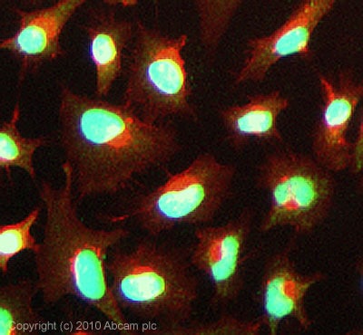
ICC/IF image of ab85555 stained HeLa cells. The cells were 100% Methanol fixed (5 min) and then incubated in 1%BSA / 10% normal Goat serum / 0.3M glycine in 0.1% PBS-Tween for 1h to permeabilise the cells and block non-specific protein-protein interactions. The cells were then incubated with the antibody (ab85555, 5µg/ml) overnight at +4°C. The secondary antibody (green) was Alexa Fluor® 488 Goat anti-Rabbit IgG (H+L) used at a 1/1000 dilution for 1h. Alexa Fluor® 594 WGA was used to label plasma membranes (red) at a 1/200 dilution for 1h. DAPI was used to stain the cell nuclei (blue) at a concentration of 1.43µM. This antibody also gave a positive result in 100% Methanol fixed (5 min) HepG2 cells at 5µg/ml, and in 4% PFA fixed (10 min) HeLa, and HepG2 cells at 5µg/ml.
-
ab85555 staining PKM2 in HeLa cells treated with resveratrol (ab120726), by ICC/IF. Decrease in PKM2 expression correlates with increased concentration of resveratrol as described in literature.
The cells were incubated at 37°C for 48h in media containing different concentrations of ab120726 (resveratrol) in DMSO, fixed with 4% formaldehyde for 10 minutes at room temperature and blocked with PBS containing 10% goat serum, 0.3 M glycine, 1% BSA and 0.1% tween for 2h at room temperature. Staining of the treated cells with ab85555 (5 µg/ml) was performed overnight at 4°C in PBS containing 1% BSA and 0.1% tween. A DyLight 488 goat anti-rabbit polyclonal antibody (ab96899) at 1/250 dilution was used as the secondary antibody. Nuclei were counterstained with DAPI and are shown in blue.
Protocols
References (1)
ab85555 has been referenced in 1 publication.
- Dai J et al. Primary prostate cancer educates bone stroma through exosomal pyruvate kinase M2 to promote bone metastasis. J Exp Med 216:2883-2899 (2019). PubMed: 31548301
Images
-
All lanes : Anti-PKM2 antibody (ab85555) at 1 µg/ml
Lane 1 : HeLa (Human epithelial carcinoma cell line) Whole Cell Lysate
Lane 2 : MEL-1 (Human embryonic stem cell, male cell line) Whole Cell Lysate (ab27198)
Lane 3 : HepG2 (Human hepatocellular liver carcinoma cell line) Whole Cell Lysate
Lane 4 : MCF7 (Human breast adenocarcinoma cell line) Whole Cell Lysate
Lane 5 : Caco 2 (Human colonic carcinoma cell line) Whole Cell Lysate
Lane 6 : SHSY-5Y (Human neuroblastoma cell line) Whole Cell Lysate
Lane 7 : Raji (Human Burkitt's lymphoma cell line) Whole Cell Lysate
Lysates/proteins at 10 µg per lane.
Secondary
All lanes : Goat polyclonal to Rabbit IgG - H&L - Pre-Adsorbed (HRP) at 1/3000 dilution
Developed using the ECL technique.
Performed under reducing conditions.
Predicted band size: 57 kDa
Observed band size: 57 kDa
-
ICC/IF image of ab85555 stained HeLa cells. The cells were 100% Methanol fixed (5 min) and then incubated in 1%BSA / 10% normal Goat serum / 0.3M glycine in 0.1% PBS-Tween for 1h to permeabilise the cells and block non-specific protein-protein interactions. The cells were then incubated with the antibody (ab85555, 5µg/ml) overnight at +4°C. The secondary antibody (green) was Alexa Fluor® 488 Goat anti-Rabbit IgG (H+L) used at a 1/1000 dilution for 1h. Alexa Fluor® 594 WGA was used to label plasma membranes (red) at a 1/200 dilution for 1h. DAPI was used to stain the cell nuclei (blue) at a concentration of 1.43µM. This antibody also gave a positive result in 100% Methanol fixed (5 min) HepG2 cells at 5µg/ml, and in 4% PFA fixed (10 min) HeLa, and HepG2 cells at 5µg/ml.
-
ab85555 staining PKM2 in HeLa cells treated with resveratrol (ab120726), by ICC/IF. Decrease in PKM2 expression correlates with increased concentration of resveratrol as described in literature.
The cells were incubated at 37°C for 48h in media containing different concentrations of ab120726 (resveratrol) in DMSO, fixed with 4% formaldehyde for 10 minutes at room temperature and blocked with PBS containing 10% goat serum, 0.3 M glycine, 1% BSA and 0.1% tween for 2h at room temperature. Staining of the treated cells with ab85555 (5 µg/ml) was performed overnight at 4°C in PBS containing 1% BSA and 0.1% tween. A DyLight 488 goat anti-rabbit polyclonal antibody (ab96899) at 1/250 dilution was used as the secondary antibody. Nuclei were counterstained with DAPI and are shown in blue.


