Anti-PICK1 antibody (ab3420)
")
Key features and details
- Rabbit polyclonal to PICK1
- Suitable for: ICC, WB, IHC-P
- Reacts with: Mouse, Rat, Human
- Isotype: IgG
Overview
-
Product name
Anti-PICK1 antibody
See all PICK1 primary antibodies -
Description
Rabbit polyclonal to PICK1 -
Host species
Rabbit -
Specificity
Detects protein interacting with Protein Kinase C (PICK 1). By Western blot, this antibody detects an ~52 kDa protein representing PICK 1 from AtT20 cell extract. Immunocytochemical staining of CV1 cells transfected with PICK 1 results in a diffuse staining pattern when expressed alone and coclustered when coexpressed with mGluR7. -
Tested applications
Suitable for: ICC, WB, IHC-Pmore details -
Species reactivity
Reacts with: Mouse, Rat, Human
Predicted to work with: Cow, Cynomolgus monkey
-
Immunogen
Synthetic peptide corresponding to Mouse PICK1 aa 1-18.
Sequence:MFADLDYDIEEDKLGIPT
(Peptide available asab4964)


Properties
-
Form
Liquid -
Storage instructions
Shipped at 4°C. Store at +4°C short term (1-2 weeks). Upon delivery aliquot. Store at -20°C or -80°C. Avoid freeze / thaw cycle. -
Storage buffer
Constituents: 0.1% BSA, 99% PBS -
 Concentration information loading...
 -
Purity
Immunogen affinity purified -
Clonality
Polyclonal -
Isotype
IgG -
Research areas
Images
-
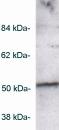
Western blot - Anti-PICK1 antibody (ab3420) This image is courtesy of an anonymous abreview.Lane 1 : An myc antibody in PBST for 16 hours at 4°C
Lane 2 : Anti-PICK1 antibody (ab3420) at 1/500 dilution (in PBST for 16 hours at 4°C)
All lanes : Transfected CHO cells expressing human Pick1 tagged with myc
Lysates/proteins at 20 µg per lane.
Secondary
Lane 2 : An HRP-conjugated goat anti-rabbit IgG polyclonal at 1/10000 dilution
Developed using the ECL technique.
Performed under reducing conditions.
Observed band size: ~52 kDa why is the actual band size different from the predicted?
Additional bands at: ~40 kDa. We are unsure as to the identity of these extra bands.
Exposure time: 2 minutes
Blocking Step: 5% Milk for 1 hour at 20°C -
 Immunocytochemistry - Anti-PICK1 antibody (ab3420) This image is courtesy of an anonymous abreview.
ab3420 staining PICK1 in mouse Hippocampal neurons by Immunocytochemistry/ Immunofluorescence. Cells were fixed in paraformaldehyde and permeabilized in 0.1% saponin/PBS prior to blocking in 3% BSA for 1 hour at room temperature. The primary antibody was diluted 1/300 in 3% BSA, 0.1% saponin /PBS and incubated with the sample for 18 hours at 4 °C. The secondary antibody was Alexa Fluor® 455-conjugated Donkey anti-Rabbit polyclonal, diluted 1/1000.
-
 Immunohistochemistry (Formalin/PFA-fixed paraffin-embedded sections) - Anti-PICK1 antibody (ab3420)IHC image of ab3420 staining in rat adult brain formalin fixed paraffin embedded tissue section, performed on a Leica BondTM system using the standard protocol F. The section was pre-treated using heat mediated antigen retrieval with sodium citrate buffer (pH6, epitope retrieval solution 1) for 20 mins. The section was then incubated with ab3420, 1µg/ml, for 15 mins at room temperature and detected using an HRP conjugated compact polymer system. DAB was used as the chromogen. The section was then counterstained with haematoxylin and mounted with DPX.
For other IHC staining systems (automated and non-automated) customers should optimize variable parameters such as antigen retrieval conditions, primary antibody concentration and antibody incubation times.
-
 Immunocytochemistry - Anti-PICK1 antibody (ab3420) This image is courtesy of an anonymous abreview.ab3420 staining PICK1 in CHO cells stably transfected with Human PICK1 by Immunocytochemistry/ Immunofluorescence. Cells were fixed in paraformaldehyde and permeabilized in 0.5% Triton X-100 prior to blocking in 10% serum for 1 hour at room temperature. The primary antibody was diluted 1/1000 in PBS, 0.5% Triton, 10% FCS and incubated with the sample for 2 hours at °C. The secondary antibody was Alexa Fluor® 488-conjugated Goat anti-Rabbit polyclonal, diluted 1/500.
 -
 Western blot - Anti-PICK1 antibody (ab3420)
Western blot of PICK1 on AtT20 cell lysate using ab3420.


