Anti-pan-AKT (phospho T308) antibody (ab8933)
 antibody (ab8933)")
Key features and details
- Rabbit polyclonal to pan-AKT (phospho T308)
- Suitable for: IHC-P, WB, Dot blot
- Reacts with: Human, Recombinant fragment
- Isotype: IgG
Overview
-
Product name
Anti-pan-AKT (phospho T308) antibody
See all pan-AKT primary antibodies -
Description
Rabbit polyclonal to pan-AKT (phospho T308) -
Host species
Rabbit -
Specificity
This antibody was made against a peptide directed against the phosphorylated form of AKT1 at T308, but due to a high degree of homology it is predicted to cross react with AKT2 and AKT3 if they are phosphorylated at the corresponding residue. Weak cross reactivity with AKT2.
-
Tested applications
Suitable for: IHC-P, WB, Dot blotmore details -
Species reactivity
Reacts with: Human, Recombinant fragment
Predicted to work with: Mouse, Rat
-
Immunogen
Synthetic peptide. This information is considered to be commercially sensitive.
-
Positive control
- IHC-P: Human lung, testis and breast tissue. WB: Human spleen, small intestine, placenta, skeletal muscle, lung, tonsil and thymus tissue lysate. GST-tagged AKT1 recombinant protein. AKT1 and AKT3 recombinant protein.
Properties
-
Form
Liquid -
Storage instructions
Shipped at 4°C. Upon delivery aliquot and store at -20°C or -80°C. Avoid repeated freeze / thaw cycles. -
Storage buffer
Preservative: 0.01% Sodium azide
Constituents: 0.42% Potassium phosphate, 0.87% Sodium chloride -
 Concentration information loading...
 -
Purity
Immunogen affinity purified -
Purification notes
This product was prepared from monospecific antiserum by immunoaffinity chromatography using phospho peptide coupled to agarose beads followed by solid phase adsorption(s) against non-phospho peptide and non-specific peptide to remove any unwanted reactivities. Assay by immunoelectrophoresis resulted in a single precipitin arc against anti-Rabbit Serum. -
Clonality
Polyclonal -
Isotype
IgG -
Research areas
Images
-
Immunohistochemistry (Formalin/PFA-fixed paraffin-embedded sections) - Anti-pan-AKT (phospho T308) antibody (ab8933)
Panel A: ab8933 (1/200 dilution, 30mins at RT) staining AKT (phospho T308) in human testis tissue in immunohistochemical analysis. Secondary used was an anti-Rabbit polyclonal HRP conjugate (Ready to use) DAB staining. Heat induced antigen retrieval was performed using Leica Bond Epitope Retrieval Buffer 1 (Citrate solution, pH6.0) for 20 minutes (ER1(20)). Counterstain is hemotoxylin.
Panel B: Secondary antibody only.
-
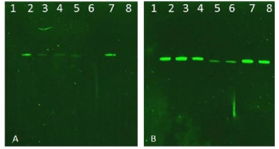 Western blot - Anti-pan-AKT (phospho T308) antibody (ab8933)All lanes : Anti-pan-AKT (phospho T308) antibody (ab8933) at 1/2270 dilution
Lane 1 : MW Protein ladder
Lane 2 : Recombinant AKT1 protein, 50ng
Lane 3 : Recombinant AKT1 protein, 50ng (phosphatase treated)
Lane 4 : Recombinant AKT1 T308A/S473A mutant protein, 50ng
Lane 5 : Recombinant AKT2 protein, 50ng
Lane 6 : Recombinant AKT2 protein, 50ng (phosphatase treated)
Lane 7 : Recombinant AKT3 protein, 50ng
Lane 8 : Recombinant AKT3 protein, 50ng (phosphatase treated)
Predicted band size: 56 kDaBlot A: ab8933 used at 1/2270.
Blot B: Anti-Akt used 1/1000.
-
 Dot Blot - Anti-pan-AKT (phospho T308) antibody (ab8933)
Dot Blot - Anti-pan-AKT (phospho T308) antibody (ab8933, 5 µg/ml).
Secondary antibody is an anti-rabbit IgG HRP used at a 1/70,000 dilution.
Exposure time 60 secs.
Columns 1 – 5, Left to Right 100, 33.33, 11.11, 3.70, 1.23 ng
Row A: AKT1-BSA peptide
Row B: AKT1 pT308 – BSA peptide
Row C: AKT1 S473 – BSA peptide
Row D: AKT1 pS473 – BSA peptide
Row E: CDC27 T244 -BSA peptide
Row F: CDC27 pT244 – BSA peptide
Row G: BSA control -
 Immunohistochemistry (Formalin/PFA-fixed paraffin-embedded sections) - Anti-pan-AKT (phospho T308) antibody (ab8933)
ab8933 (4µg/ml) staining AKT (phospho T308) in human breast using an automated system (DAKO Autostainer Plus). Using this protocol there is strong staining of membrane and cytoplasmic compartment within the breast ductal regions.
Sections were rehydrated and antigen retrieved with the Dako 3 in 1 AR buffer EDTA pH 9.0 in a DAKO PT link. Slides were peroxidase blocked in 3% H2O2 in methanol for 10 mins. They were then blocked with Dako Protein block for 10 minutes (containing casein 0.25% in PBS) then incubated with primary antibody for 20 min and detected with Dako envision flex amplification kit for 30 minutes. Colorimetric detection was completed with Diaminobenzidine for 5 minutes. Slides were counterstained with Haematoxylin and coverslipped under DePeX. Please note that, for manual staining, optimization of primary antibody concentration and incubation time is recommended. Signal amplification may be required. -
 Immunohistochemistry (Formalin/PFA-fixed paraffin-embedded sections) - Anti-pan-AKT (phospho T308) antibody (ab8933)
ab8933 (1/200 dilution, 30mins at RT) staining AKT (phospho T308) in human lung tissue in immunohistochemical analysis. Secondary used was an anti-Rabbit polyclonal HRP conjugate (Ready to use, 8mins at RT) DAB staining. HIER using citrate buffer for 20mins. Counterstain is hemotoxylin.
-
 Immunohistochemistry (Formalin/PFA-fixed paraffin-embedded sections) - Anti-pan-AKT (phospho T308) antibody (ab8933)
ab8933 (1/200 dilution, 30mins at RT) staining AKT (phospho T308) in human lymph node in breast tissue in immunohistochemical analysis. Secondary used was an anti-Rabbit polyclonal HRP conjugate (Ready to use, 8mins at RT) DAB staining. HIER using citrate buffer for 20mins. Counterstain is hemotoxylin.
-
 Western blot - Anti-pan-AKT (phospho T308) antibody (ab8933)All lanes : Anti-pan-AKT (phospho T308) antibody (ab8933) at 1/1000 dilution (overnight at 4degC)
Lane 1 : GST-tagged AKT1 recombinant protein, 50ng
Lane 2 : GST-tagged AKT1 active recombinant protein, 50ng
Secondary
All lanes : DyLight™ 649 rabbit secondary antibody, (30mins at RT) at 1/20000 dilution
Predicted band size: 56 kDaRecombinant protein expected to run ~80-100 kDa.
-
 Western blot - Anti-pan-AKT (phospho T308) antibody (ab8933)All lanes : Anti-pan-AKT (phospho T308) antibody (ab8933) at 1/1000 dilution
Lane 1 : Human spleen whole tissue lysate
Lane 2 : Human small intestine whole tissue lysate
Lane 3 : Human placenta whole tissue lysate
Lane 4 : Human skeletal muscle whole tissue lysate
Lane 5 : Human brain cerebellum whole tissue lysate
Lane 6 : Human lung whole tissue lysate
Lane 7 : Human tonsil whole tissue lysate
Lane 8 : Human thymus whole tissue lysate
Secondary
All lanes : Goat anti-Rabbit Ig HRP at 1/40000 dilution
Predicted band size: 56 kDa
Exposure time: 8 seconds


