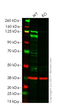
Anti-Notch1 antibody (ab65297)
Images
-
All lanes : Anti-Notch1 antibody (ab65297) at 1 µg/ml
Lane 1 : Wild-type HeLa cell lysate
Lane 2 : NOTCH1 knockout HeLa cell lysate
Lysates/proteins at 20 µg per lane.
Performed under reducing conditions.
Predicted band size: 272 kDa
Observed band size: 110 kDa why is the actual band size different from the predicted?Lanes 1- 2: Merged signal (red and green). Green - ab65297 observed at 110 kDa. Red - Anti-GAPDH antibody [6C5] - Loading Control (ab8245) observed at 37 kDa.
ab65297 was shown to react with Notch1 in wild-type HeLa cells in western blot. Loss of signal was observed when knockout cell line ab261762 (knockout cell lysate ab257006) was used. Wild-type HeLa and NOTCH1 knockout HeLa cell lysates were subjected to SDS-PAGE. Membrane was blocked for 1 hour at room temperature in 0.1% TBST with 3% non-fat dried milk. ab65297 and Anti-GAPDH antibody [6C5] - Loading Control (ab8245) overnight at 4°C at a 1 µg/ml and a 1 in 20000 dilution respectively. Blots were developed with Goat anti-Rabbit IgG H&L (IRDye®800CW) preadsorbed (ab216773) and Goat anti-Mouse IgG H&L (IRDye®680RD) preadsorbed (ab216776) secondary antibodies at 1 in 20000 dilution for 1 hour at room temperature before imaging.
-
All lanes : Anti-Notch1 antibody (ab65297) at 1 µg/ml
Lane 1 : Wild-type HAP1 whole cell lysate
Lane 2 : NOTCH1 knockout HAP1 whole cell lysate
Lysates/proteins at 40 µg per lane.
Predicted band size: 272 kDaLanes 1 - 2: Merged signal (red and green). Green - ab65297 observed at 110 kDa. Red - loading control, ab8245, observed at 37 kDa.
ab65297 was shown to recognize in wild-type HAP1 cells as signal was lost at the expected MW in NOTCH1 knockout cells. Additional cross-reactive bands were observed in the wild-type and knockout cells. Wild-type and NOTCH1 knockout samples were subjected to SDS-PAGE. The membrane was blocked with 3% NF Milk. ab65297 and ab8245 (Mouse anti GAPDH loading control) were incubated overnight at 4°C at 1 ug/ml and 1/20000 dilution respectively. Blots were developed with Goat anti-Rabbit IgG H&L (IRDye® 800CW) preabsorbed ab216773 and Goat anti-Mouse IgG H&L (IRDye® 680RD) preabsorbed ab216776 secondary antibodies at 1/20000 dilution for 1 hour at room temperature before imaging.
-
All lanes : ab65927 at 1 mg/ml
Lane 1 : Human HEK293 whole cell lysate
Lane 2 : Human SKNSH whole cell lysate
Lane 3 : Human Jurkat whole cell lysate
Lysates/proteins at 10 µg per lane.
Secondary
All lanes : Goat polyclonal to Rabbit IgG - H&L - Pre-Adsorbed (HRP) at 1/3000 dilution
Developed using the ECL technique.
Performed under reducing conditions.
Predicted band size: 272 kDa
Observed band size: 112 kDa why is the actual band size different from the predicted?
Exposure time: 8 minutes
-
Notch1 was immunoprecipitated using 0.5mg Hek293 whole cell extract, 5µg of Rabbit polyclonal to Notch1 and 50µl of protein G magnetic beads (+). No antibody was added to the control (-).
The antibody was incubated under agitation with Protein G beads for 10min, Hek293 whole cell extract lysate diluted in RIPA buffer was added to each sample and incubated for a further 10min under agitation.
Proteins were eluted by addition of 40µl SDS loading buffer and incubated for 10min at 70°C; 10µl of each sample was separated on a SDS PAGE gel, transferred to a nitrocellulose membrane, blocked with 5% BSA and probed with ab65297.
Secondary: Mouse monoclonal [SB62a] Secondary Antibody to Rabbit IgG light chain (HRP) (ab99697).
Band: 120kDa; Notch1


