Anti-Myeloperoxidase antibody (ab65871)
")
Key features and details
- Rabbit polyclonal to Myeloperoxidase
- Suitable for: WB, IHC-P
- Reacts with: Mouse, Rat, Human
- Isotype: IgG
Overview
-
Product name
Anti-Myeloperoxidase antibody
See all Myeloperoxidase primary antibodies -
Description
Rabbit polyclonal to Myeloperoxidase -
Host species
Rabbit -
Tested applications
Suitable for: WB, IHC-Pmore details -
Species reactivity
Reacts with: Mouse, Rat, Human -
Immunogen
Synthetic peptide corresponding to Human Myeloperoxidase (C terminal).
Database link: P05164 -
Positive control
- IHC-P: Human colon, tonsil, ovary tissue, liver cancer tissue; rat colon tissue; mouse spleen tissue. WB: Rat brain tissue.
-
General notes
Reproducibility is key to advancing scientific discovery and accelerating scientists’ next breakthrough.
Abcam is leading the way with our range of recombinant antibodies, knockout-validated antibodies and knockout cell lines, all of which support improved reproducibility.
We are also planning to innovate the way in which we present recommended applications and species on our product datasheets, so that only applications & species that have been tested in our own labs, our suppliers or by selected trusted collaborators are covered by our Abpromise™ guarantee.
In preparation for this, we have started to update the applications & species that this product is Abpromise guaranteed for.
We are also updating the applications & species that this product has been “predicted to work with,” however this information is not covered by our Abpromise guarantee.
Applications & species from publications and Abreviews that have not been tested in our own labs or in those of our suppliers are not covered by the Abpromise guarantee.
Please check that this product meets your needs before purchasing. If you have any questions, special requirements or concerns, please send us an inquiry and/or contact our Support team ahead of purchase. Recommended alternatives for this product can be found below, as well as customer reviews and Q&As.
Properties
-
Form
Lyophilized:Add 0.2ml of distilled water to yield a dilution of 500 ug/mL -
Storage instructions
Shipped at 4°C. Upon delivery aliquot and store at -20°C or -80°C. Avoid repeated freeze / thaw cycles. -
Storage buffer
Preservatives: 0.025% Thimerosal (merthiolate), 0.025% Sodium azide
Constituents: 2.5% BSA, 0.45% Sodium chloride, 0.1% Dibasic monohydrogen sodium phosphate -
 Concentration information loading...
 -
Purity
Immunogen affinity purified -
Clonality
Polyclonal -
Isotype
IgG -
Research areas
Images
-
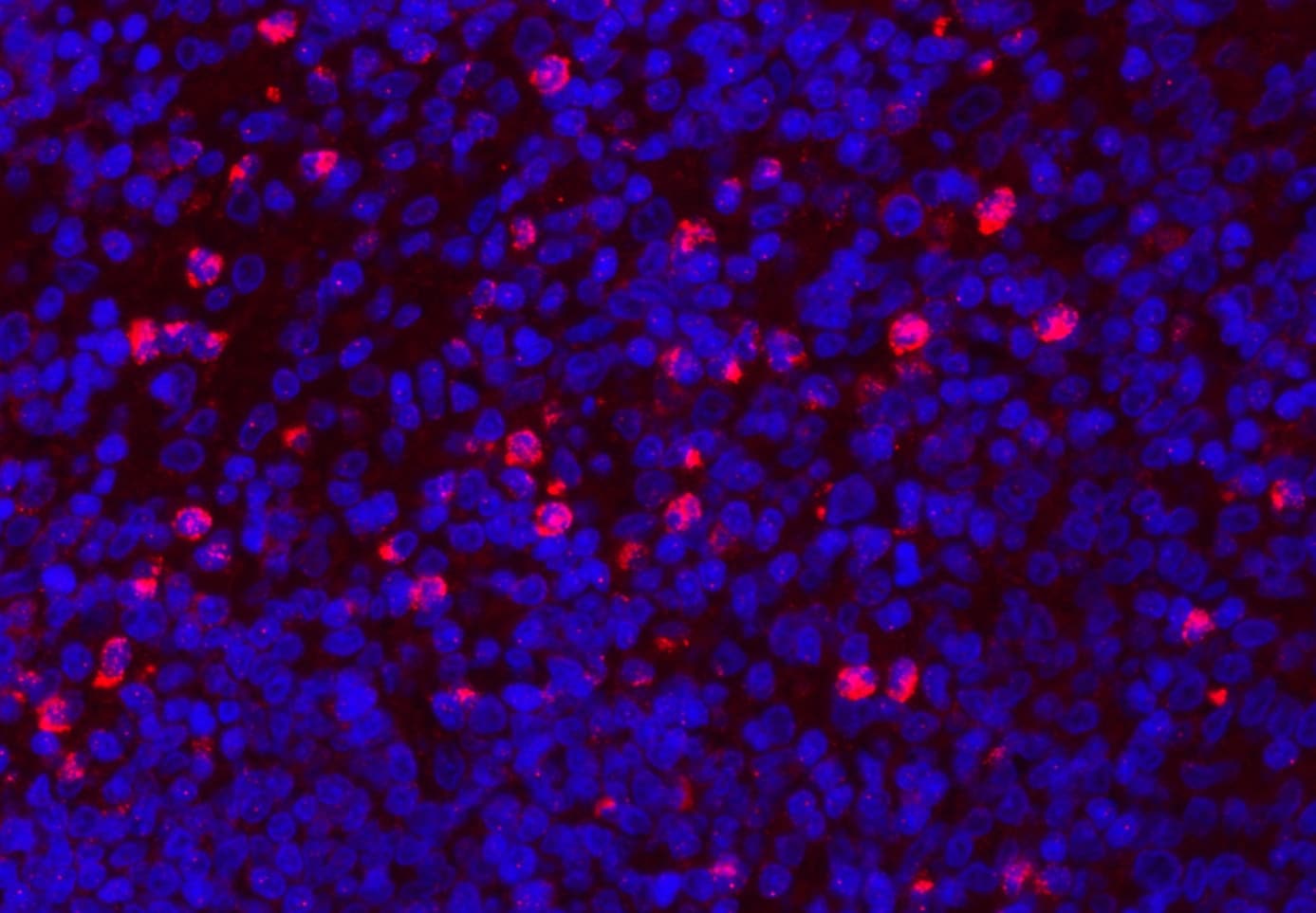
Immunohistochemistry (Formalin/PFA-fixed paraffin-embedded sections) - Anti-Myeloperoxidase antibody - Carboxyterminal end (ab65871)
Immunohistochemistry (Formalin/PFA-fixed paraffin-embedded sections) analysis of Myeloperoxidase using ab65871.
Myeloperoxidase was detected in paraffin-embedded section of human colon tissues. Heat mediated antigen retrieval was performed in citrate buffer (pH6, epitope retrieval solution) for 20 mins. The tissue section was blocked with 10% goat serum. The tissue section was then incubated with 1μg/mL overnight at 4°C. Cy3 Conjugated Goat Anti-Rabbit IgG was used as secondary antibody at 1:100 dilution and incubated for 30 minutes at 37°C. The section was counterstained with DAPI. Visualize using a fluorescence microscope and filter sets appropriate for the label used.
-
 Western blot - Anti-Myeloperoxidase antibody - Carboxyterminal end (ab65871)Anti-Myeloperoxidase antibody (ab65871) at 2 µg/ml + rat brain tissue lysate
Predicted band size: 84 kDa
Observed band size: 170 kDa why is the actual band size different from the predicted?
-
 Immunohistochemistry (Formalin/PFA-fixed paraffin-embedded sections) - Anti-Myeloperoxidase antibody - Carboxyterminal end (ab65871)
Immunohistochemistry (Formalin/PFA-fixed paraffin-embedded sections) analysis of Myeloperoxidase using ab65871.
Myeloperoxidase was detected in paraffin-embedded section of human tonsil tissues. Heat mediated antigen retrieval was performed in citrate buffer (pH6, epitope retrieval solution ) for 20 mins. The tissue section was blocked with 10% goat serum. The tissue section was then incubated with 1μg/mL ab65871 overnight at 4°C. Cy3 Conjugated Goat Anti-Rabbit IgG was used as secondary antibody at 1:100 dilution and incubated for 30 minutes at 37°C. The section was counterstained with DAPI. Visualize using a fluorescence microscope and filter sets appropriate for the label used.
-
 Immunohistochemistry (Formalin/PFA-fixed paraffin-embedded sections) - Anti-Myeloperoxidase antibody - Carboxyterminal end (ab65871)
Immunohistochemistry (Formalin/PFA-fixed paraffin-embedded sections) analysis of Myeloperoxidase using ab65871.
Myeloperoxidase was detected in paraffin-embedded section of human liver cancer tissues. Heat mediated antigen retrieval was performed in citrate buffer (pH6, epitope retrieval solution) for 20 mins. The tissue section was blocked with 10% goat serum. The tissue section was then incubated with 1μg/ml ab65871 overnight at 4°C. Biotinylated goat anti-rabbit IgG was used as secondary antibody and incubated for 30 minutes at 37°C. The tissue section was developed using Strepavidin-Biotin-Complex with DAB as the chromogen. -
 Immunohistochemistry (Formalin/PFA-fixed paraffin-embedded sections) - Anti-Myeloperoxidase antibody - Carboxyterminal end (ab65871)
Immunohistochemistry (Formalin/PFA-fixed paraffin-embedded sections) analysis of Myeloperoxidase using ab65871.
Myeloperoxidase was detected in paraffin-embedded section of rat colon tissues. Heat mediated antigen retrieval was performed in citrate buffer (pH6, epitope retrieval solution ) for 20 mins. The tissue section was blocked with 10% goat serum. The tissue section was then incubated with 1μg/mL ab65871 overnight at 4°C. Cy3 Conjugated Goat Anti-Rabbit IgG was used as secondary antibody at 1:100 dilution and incubated for 30 minutes at 37°C. The section was counterstained with DAPI. Visualize using a fluorescence microscope and filter sets appropriate for the label used.
-
 Immunohistochemistry (Formalin/PFA-fixed paraffin-embedded sections) - Anti-Myeloperoxidase antibody - Carboxyterminal end (ab65871)
Immunohistochemistry (Formalin/PFA-fixed paraffin-embedded sections) analysis of Myeloperoxidase using ab65871.
Myeloperoxidase was detected in paraffin-embedded section of mouse spleen tissues. Heat mediated antigen retrieval was performed in citrate buffer (pH6, epitope retrieval solution ) for 20 mins. The tissue section was blocked with 10% goat serum. The tissue section was then incubated with 1μg/mL ab65871 overnight at 4°C. Cy3 Conjugated Goat Anti-Rabbit IgG was used as secondary antibody at 1:100 dilution and incubated for 30 minutes at 37°C. The section was counterstained with DAPI. Visualize using a fluorescence microscope and filter sets appropriate for the label used.
-
 Immunohistochemistry (Formalin/PFA-fixed paraffin-embedded sections) - Anti-Myeloperoxidase antibody - Carboxyterminal end (ab65871)This image shows human ovary tissue stained with ab65871 at 1µg/ml.


