Anti-MUC2 antibody (ab90007)
")
Key features and details
- Rabbit polyclonal to MUC2
- Suitable for: IHC-P, ICC/IF
- Reacts with: Human
- Isotype: IgG
Overview
-
Product name
Anti-MUC2 antibody
See all MUC2 primary antibodies -
Description
Rabbit polyclonal to MUC2 -
Host species
Rabbit -
Tested applications
Suitable for: IHC-P, ICC/IFmore details -
Species reactivity
Reacts with: Human
Predicted to work with: Mouse, Rat
-
Immunogen
-
Positive control
- This antibody gave a positive signal in human colon tissue sections. ICC/IF: HT29 cell line
-
General notes
Reproducibility is key to advancing scientific discovery and accelerating scientists’ next breakthrough.
Abcam is leading the way with our range of recombinant antibodies, knockout-validated antibodies and knockout cell lines, all of which support improved reproducibility.
We are also planning to innovate the way in which we present recommended applications and species on our product datasheets, so that only applications & species that have been tested in our own labs, our suppliers or by selected trusted collaborators are covered by our Abpromise™ guarantee.
In preparation for this, we have started to update the applications & species that this product is Abpromise guaranteed for.
We are also updating the applications & species that this product has been “predicted to work with,” however this information is not covered by our Abpromise guarantee.
Applications & species from publications and Abreviews that have not been tested in our own labs or in those of our suppliers are not covered by the Abpromise guarantee.
Please check that this product meets your needs before purchasing. If you have any questions, special requirements or concerns, please send us an inquiry and/or contact our Support team ahead of purchase. Recommended alternatives for this product can be found below, as well as customer reviews and Q&As.
Properties
-
Form
Liquid -
Storage instructions
Shipped at 4°C. Store at +4°C short term (1-2 weeks). Upon delivery aliquot. Store at -20°C or -80°C. Avoid freeze / thaw cycle. -
Storage buffer
pH: 7.40
Preservative: 0.02% Sodium azide
Constituent: PBS
Batches of this product that have a concentration Concentration information loading...
Purity
Immunogen affinity purifiedClonality
PolyclonalIsotype
IgGResearch areas
Associated products
-
Compatible Secondaries
-
Isotype control
Applications
Our Abpromise guarantee covers the use of ab90007 in the following tested applications.
The application notes include recommended starting dilutions; optimal dilutions/concentrations should be determined by the end user.
Application Abreviews Notes IHC-P Use a concentration of 5 µg/ml. Perform heat mediated antigen retrieval with citrate buffer pH 6 before commencing with IHC staining protocol. ICC/IF Use a concentration of 5 µg/ml. Target
-
Function
Coats the epithelia of the intestines, airways, and other mucus membrane-containing organs. Thought to provide a protective, lubricating barrier against particles and infectious agents at mucosal surfaces. Major constituent of both the inner and outer mucus layers of the colon and may play a role in excluding bacteria from the inner mucus layer. -
Tissue specificity
Colon, small intestine, colonic tumors, bronchus, cervix and gall bladder. -
Sequence similarities
Contains 1 CTCK (C-terminal cystine knot-like) domain.
Contains 1 TIL (trypsin inhibitory-like) domain.
Contains 2 VWFC domains.
Contains 4 VWFD domains. -
Post-translational
modificationsO-glycosylated.
May undergo proteolytic cleavage in the outer mucus layer of the colon, contributing to the expanded volume and loose nature of this layer which allows for bacterial colonization in contrast to the inner mucus layer which is dense and devoid of bacteria.
At low pH of 6 and under, undergoes autocatalytic cleavage in vitro in the N-terminal region of the fourth VWD domain. It is likely that this also occurs in vivo and is triggered by the low pH of the late secretory pathway. -
Cellular localization
Secreted. In the intestine, secreted into the inner and outer mucus layers. - Information by UniProt
-
Database links
- Entrez Gene: 4583 Human
- Entrez Gene: 17831 Mouse
- Entrez Gene: 24572 Rat
- Omim: 158370 Human
- SwissProt: Q02817 Human
- SwissProt: Q80Z19 Mouse
- SwissProt: Q62635 Rat
- Unigene: 315 Human
see all -
Alternative names
- Intestinal mucin 2 antibody
- Intestinal mucin-2 antibody
- MLP antibody
see all
Images
-
Immunohistochemistry (Formalin/PFA-fixed paraffin-embedded sections) - Anti-MUC2 antibody (ab90007)IHC image of MUC2 staining in human normal colon FFPE section, performed on a BondTM system using the standard protocol F. The section was pre-treated using heat mediated antigen retrieval with sodium citrate buffer (pH6, epitope retrieval solution 1) for 20 mins. The section was then incubated with ab90007, 5µg/ml, for 15 mins at room temperature and detected using an HRP conjugated compact polymer system. DAB was used as the chromogen. The section was then counterstained with haematoxylin and mounted with DPX
-
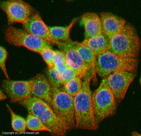 Immunocytochemistry/ Immunofluorescence - Anti-MUC2 antibody (ab90007)
ab90007 stained in HT29 cells. Cells were fixed with 100% methanol (5 min) at room temperature and incubated with PBS containing 10% goat serum, 0.3 M glycine, 1% BSA and 0.1% Triton for 1h at room temperature to permeabilise the cells and block non-specific protein-protein interactions. The cells were then incubated with the antibody ab90007 at 5µg/ml and ab7291 (Mouse monoclonal to alpha Tubulin - Loading Control) used at a 1/1000 dilution overnight at +4°C. The secondary antibodies were ab150081, Goat Anti-Rabbit IgG H&L (Alexa Fluor® 488) preadsorbed, (pseudo-colored green) and ab150120, Goat polyclonal Secondary Antibody to Mouse IgG - H&L (Alexa Fluor® 594) preadsorbed, (colored red), both used at a 1/1000 dilution for 1 hour at room temperature. DAPI was used to stain the cell nuclei (colored blue) at a concentration of 1.43 µM for 1hour at room temperature.
Protocols
Datasheets and documents
References (2)
ab90007 has been referenced in 2 publications.
- Choi W et al. YAP/TAZ Initiates Gastric Tumorigenesis via Upregulation of MYC. Cancer Res 78:3306-3320 (2018). PubMed: 29669762
- Shen C et al. Deoxycholic acid (DCA) confers an intestinal phenotype on esophageal squamous epithelium via induction of the stemness-associated reprogramming factors OCT4 and SOX2. Cell Cycle 15:1439-49 (2016). PubMed: 27096226
Images
-
Immunohistochemistry (Formalin/PFA-fixed paraffin-embedded sections) - Anti-MUC2 antibody (ab90007)IHC image of MUC2 staining in human normal colon FFPE section, performed on a BondTM system using the standard protocol F. The section was pre-treated using heat mediated antigen retrieval with sodium citrate buffer (pH6, epitope retrieval solution 1) for 20 mins. The section was then incubated with ab90007, 5µg/ml, for 15 mins at room temperature and detected using an HRP conjugated compact polymer system. DAB was used as the chromogen. The section was then counterstained with haematoxylin and mounted with DPX
-
Immunocytochemistry/ Immunofluorescence - Anti-MUC2 antibody (ab90007)
ab90007 stained in HT29 cells. Cells were fixed with 100% methanol (5 min) at room temperature and incubated with PBS containing 10% goat serum, 0.3 M glycine, 1% BSA and 0.1% Triton for 1h at room temperature to permeabilise the cells and block non-specific protein-protein interactions. The cells were then incubated with the antibody ab90007 at 5µg/ml and ab7291 (Mouse monoclonal to alpha Tubulin - Loading Control) used at a 1/1000 dilution overnight at +4°C. The secondary antibodies were ab150081, Goat Anti-Rabbit IgG H&L (Alexa Fluor® 488) preadsorbed, (pseudo-colored green) and ab150120, Goat polyclonal Secondary Antibody to Mouse IgG - H&L (Alexa Fluor® 594) preadsorbed, (colored red), both used at a 1/1000 dilution for 1 hour at room temperature. DAPI was used to stain the cell nuclei (colored blue) at a concentration of 1.43 µM for 1hour at room temperature.


