Anti-MSY2/YBOX2/YBX2 antibody (ab33164)
Key features and details
- Rabbit polyclonal to MSY2/YBOX2/YBX2
- Suitable for: ICC/IF, IHC-P, WB
- Reacts with: Mouse, Human, Common marmoset
- Isotype: IgG
Overview
-
Product name
Anti-MSY2/YBOX2/YBX2 antibody
See all MSY2/YBOX2/YBX2 primary antibodies -
Description
Rabbit polyclonal to MSY2/YBOX2/YBX2 -
Host species
Rabbit -
Tested applications
Suitable for: ICC/IF, IHC-P, WBmore details -
Species reactivity
Reacts with: Mouse, Human, Common marmoset -
Immunogen
Synthetic peptide corresponding to Mouse MSY2/YBOX2/YBX2 aa 300 to the C-terminus (C terminal).
(Peptide available asab27460) -
Positive control
- This antibody gave a positive signal in Human normal testis FFPE section. This antibody gave a positive result when used in the following formaldehyde fixed cell lines: F9.
-
General notes
This product was previously labelled as MSY2/YBOX2
Properties
-
Form
Liquid -
Storage instructions
Shipped at 4°C. Store at +4°C short term (1-2 weeks). Upon delivery aliquot. Store at -20°C or -80°C. Avoid freeze / thaw cycle. -
Storage buffer
pH: 7.40
Preservative: 0.02% Sodium azide
Constituent: PBS
Batches of this product that have a concentration Concentration information loading...
Purity
Immunogen affinity purifiedClonality
PolyclonalIsotype
IgGResearch areas
Associated products
-
Compatible Secondaries
-
Isotype control
-
Recombinant Protein
Applications
Our Abpromise guarantee covers the use of ab33164 in the following tested applications.
The application notes include recommended starting dilutions; optimal dilutions/concentrations should be determined by the end user.
Application Abreviews Notes ICC/IF Use a concentration of 5 µg/ml. IHC-P Use a concentration of 1 mg/ml. Perform heat mediated antigen retrieval before commencing with IHC staining protocol. WB Use at an assay dependent concentration. Predicted molecular weight: 38 kDa. Target
- Information by UniProt
-
Database links
- Entrez Gene: 51087 Human
- Omim: 611447 Human
- SwissProt: Q9Y2T7 Human
- SwissProt: Q9Z2C8 Mouse
- Unigene: 567494 Human
- Unigene: 29286 Mouse
-
Alternative names
- Contrin antibody
- CSDA 3 antibody
- CSDA3 antibody
see all
Images
-
 Immunohistochemistry (Formalin/PFA-fixed paraffin-embedded sections) - Anti-MSY2/YBOX2/YBX2 antibody (ab33164)IHC image of MSY2/BOX2 staining in human testis formalin fixed paraffin embedded tissue section, performed on a Leica BondTM system using the standard protocol F. The section was pre-treated using heat mediated antigen retrieval with sodium citrate buffer (pH6, epitope retrieval solution 1) for 20 mins. The section was then incubated with ab33164, 1µg/ml, for 15 mins at room temperature and detected using an HRP conjugated compact polymer system. DAB was used as the chromogen. The section was then counterstained with haematoxylin and mounted with DPX.
 -
ICC/IF image of ab33164 stained F9 cells. The cells were 4% formaldehyde fixed (10 min) and then incubated in 1%BSA / 10% normal goat serum / 0.3M glycine in 0.1% PBS-Tween for 1h to permeabilise the cells and block non-specific protein-protein interactions. The cells were then incubated with the antibody ab33164 at 5µg/ml overnight at +4°C. The secondary antibody (green) was DyLight® 488 goat anti- rabbit (ab96899) IgG (H+L) used at a 1/250 dilution for 1h. Alexa Fluor® 594 WGA was used to label plasma membranes (red) at a 1/200 dilution for 1h. DAPI was used to stain the cell nuclei (blue) at a concentration of 1.43µM.
-
 Immunohistochemistry (Formalin/PFA-fixed paraffin-embedded sections) - Anti-MSY2/YBOX2/YBX2 antibody (ab33164)Image courtesy of Human Protein Atlas
Image courtesy of Human Protein Atlas
ab33164 staining MSY2/YBOX2/YBX2 in human testis tissue, showing staining in the seminiferous tubules. Paraffin embedded human skin tissue was incubated with ab33164 (1/200 dilution) for 30 mins at room temperature. Antigen retrieval was performed by heat induction in citrate buffer pH 6.
ab33164 was tested in a tissue microarray (TMA) containing a wide range of normal and cancer tissues as well as a cell microarray consisting of a range of commonly used, well characterised human cell lines. Further results for this antibody can be found at www.proteinatlas.org
-
All lanes : Anti-MSY2/YBOX2/YBX2 antibody (ab33164) at 1 µg/ml
Lane 1 : MEL-2 (Human embryonic stem cell, female cell line) Whole Cell Lysate (ab27196)
Lane 2 : F9 (Mouse embryonic carcinoma cell line) Whole Cell Lysate (ab27193)
Lane 3 : Embryonic Germ Cell Lysate
Lysates/proteins at 20 µg per lane.
Secondary
All lanes : Rabbit IgG secondary antibody (ab28446) at 1/10000 dilution
Predicted band size: 38 kDa
Observed band size: 37 kDa why is the actual band size different from the predicted?
Additional bands at: 39 kDa (possible glycosylated form), 39 kDa (possible post-translational modification), 42 kDa (possible glycosylated form), 42 kDa (possible post-translational modification)
This antibody recognised a number of bands included 3 clustered around the YBOX2 predicted molecular weight of 38 kDa. We cannot be sure which of these bands, if any, corresponds to YBOX2. We are also unsure whether YBOX2 would be expected to be expressed at a detectable level for Western Blotting in the lysates tested. -
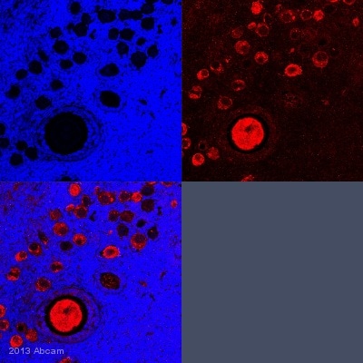
 Immunohistochemistry (Formalin/PFA-fixed paraffin-embedded sections) - Anti-MSY2/YBOX2/YBX2 antibody (ab33164)This image is courtesy of an Abreview from Zachary Yu-Ching Lin.
IHC-P image of MSY2/YBOX2/YBX2 staining with ab33164 on tissue sections from adult marmoset ovary. The sections were subjected to heat-mediated antigen retrieval using Dako antigen retrieval solution. The sections were then blocked with 5% milk for 30 minutes at 25°C, before incubation with ab33164 (1/100 dilution) for 18 hours at 4°C. The secondary was an Alexa-Fluor 555 conjugated goat anti-rabbit polyclonal, used at a 1/500 dilution.
Protocols
Datasheets and documents
References (3)
ab33164 has been referenced in 3 publications.
- Mihola O et al. Histone methyltransferase PRDM9 is not essential for meiosis in male mice. Genome Res 29:1078-1086 (2019). PubMed: 31186301
- Woods DC & Tilly JL Isolation, characterization and propagation of mitotically active germ cells from adult mouse and human ovaries. Nat Protoc 8:966-88 (2013). PubMed: 23598447
- White YA et al. Oocyte formation by mitotically active germ cells purified from ovaries of reproductive-age women. Nat Med 18:413-21 (2012). PubMed: 22366948
Images
-
 Immunohistochemistry (Formalin/PFA-fixed paraffin-embedded sections) - Anti-MSY2/YBOX2/YBX2 antibody (ab33164)IHC image of MSY2/BOX2 staining in human testis formalin fixed paraffin embedded tissue section, performed on a Leica BondTM system using the standard protocol F. The section was pre-treated using heat mediated antigen retrieval with sodium citrate buffer (pH6, epitope retrieval solution 1) for 20 mins. The section was then incubated with ab33164, 1µg/ml, for 15 mins at room temperature and detected using an HRP conjugated compact polymer system. DAB was used as the chromogen. The section was then counterstained with haematoxylin and mounted with DPX.
 -
ICC/IF image of ab33164 stained F9 cells. The cells were 4% formaldehyde fixed (10 min) and then incubated in 1%BSA / 10% normal goat serum / 0.3M glycine in 0.1% PBS-Tween for 1h to permeabilise the cells and block non-specific protein-protein interactions. The cells were then incubated with the antibody ab33164 at 5µg/ml overnight at +4°C. The secondary antibody (green) was DyLight® 488 goat anti- rabbit (ab96899) IgG (H+L) used at a 1/250 dilution for 1h. Alexa Fluor® 594 WGA was used to label plasma membranes (red) at a 1/200 dilution for 1h. DAPI was used to stain the cell nuclei (blue) at a concentration of 1.43µM.
-
 Immunohistochemistry (Formalin/PFA-fixed paraffin-embedded sections) - Anti-MSY2/YBOX2/YBX2 antibody (ab33164) Image courtesy of Human Protein Atlas
Image courtesy of Human Protein Atlas
ab33164 staining MSY2/YBOX2/YBX2 in human testis tissue, showing staining in the seminiferous tubules. Paraffin embedded human skin tissue was incubated with ab33164 (1/200 dilution) for 30 mins at room temperature. Antigen retrieval was performed by heat induction in citrate buffer pH 6.
ab33164 was tested in a tissue microarray (TMA) containing a wide range of normal and cancer tissues as well as a cell microarray consisting of a range of commonly used, well characterised human cell lines. Further results for this antibody can be found at www.proteinatlas.org
-
All lanes : Anti-MSY2/YBOX2/YBX2 antibody (ab33164) at 1 µg/ml
Lane 1 : MEL-2 (Human embryonic stem cell, female cell line) Whole Cell Lysate (ab27196)
Lane 2 : F9 (Mouse embryonic carcinoma cell line) Whole Cell Lysate (ab27193)
Lane 3 : Embryonic Germ Cell Lysate
Lysates/proteins at 20 µg per lane.
Secondary
All lanes : Rabbit IgG secondary antibody (ab28446) at 1/10000 dilution
Predicted band size: 38 kDa
Observed band size: 37 kDa why is the actual band size different from the predicted?
Additional bands at: 39 kDa (possible glycosylated form), 39 kDa (possible post-translational modification), 42 kDa (possible glycosylated form), 42 kDa (possible post-translational modification)
This antibody recognised a number of bands included 3 clustered around the YBOX2 predicted molecular weight of 38 kDa. We cannot be sure which of these bands, if any, corresponds to YBOX2. We are also unsure whether YBOX2 would be expected to be expressed at a detectable level for Western Blotting in the lysates tested. -
 Immunohistochemistry (Formalin/PFA-fixed paraffin-embedded sections) - Anti-MSY2/YBOX2/YBX2 antibody (ab33164) This image is courtesy of an Abreview from Zachary Yu-Ching Lin.
IHC-P image of MSY2/YBOX2/YBX2 staining with ab33164 on tissue sections from adult marmoset ovary. The sections were subjected to heat-mediated antigen retrieval using Dako antigen retrieval solution. The sections were then blocked with 5% milk for 30 minutes at 25°C, before incubation with ab33164 (1/100 dilution) for 18 hours at 4°C. The secondary was an Alexa-Fluor 555 conjugated goat anti-rabbit polyclonal, used at a 1/500 dilution.


