Anti-Lck antibody (ab3885)
")
Key features and details
- Rabbit polyclonal to Lck
- Suitable for: WB, ICC/IF
- Reacts with: Mouse, Human, African green monkey
- Isotype: IgG
Overview
-
Product name
Anti-Lck antibody
See all Lck primary antibodies -
Description
Rabbit polyclonal to Lck -
Host species
Rabbit -
Specificity
This antibody was designed to recognise the activated form of Lck and was raised for that purpose against the linker that connects the SH2 domain and the tyrosine kinase domain of the kinase. This linker sits in the SH3 substrate binding site in the inactive kinase. The position of the linker is based on the crystal structure of inactive Src. However, the antibody does not seem to distinguish the active and inactive forms of Lck. In IF, it recognises kinase dead Lck at the same concentration as constitutively active Lck. In western blot, the antibody is highly specific for Lck versus other tyrosine kinases (see blot). Even, if the antibody was specific for active Lck in IF , it would be expected to recognise both active and inactive forms in Western blot. This is because the linker that the antibody recognises is present in both active and inactive Lck, it was just expected to be concealed in native form inactive Lck. -
Tested applications
Suitable for: WB, ICC/IFmore details -
Species reactivity
Reacts with: Mouse, Human, African green monkey
Predicted to work with: Chicken
-
Immunogen
Synthetic peptide corresponding to Human Lck aa 200-300 conjugated to keyhole limpet haemocyanin.
(Peptide available asab13750) -
General notes
Lck (p56lck), a member of the Src family of non-receptor tyrosine protein kinases, is expressed predominantly in T cells.
Properties
-
Form
Liquid -
Storage instructions
Shipped at 4°C. Store at +4°C short term (1-2 weeks). Upon delivery aliquot. Store at -20°C or -80°C. Avoid freeze / thaw cycle. -
Storage buffer
pH: 7.40
Preservative: 0.02% Sodium azide
Constituent: PBS
Batches of this product that have a concentration Concentration information loading...
Purity
Immunogen affinity purifiedPrimary antibody notes
Lck (p56lck), a member of the Src family of non-receptor tyrosine protein kinases, is expressed predominantly in T cells.Clonality
PolyclonalIsotype
IgGResearch areas
Associated products
-
Compatible Secondaries
-
Isotype control
-
Recombinant Protein
Applications
Our Abpromise guarantee covers the use of ab3885 in the following tested applications.
The application notes include recommended starting dilutions; optimal dilutions/concentrations should be determined by the end user.
Application Abreviews Notes WB 1/1000. Detects a band of approximately 57 kDa (predicted molecular weight: 57 kDa). ICC/IF 1/200 - 1/500. Target
-
Function
Tyrosine kinase that plays an essential role for the selection and maturation of developing T-cell in the thymus and in mature T-cell function. Is constitutively associated with the cytoplasmic portions of the CD4 and CD8 surface receptors and plays a key role in T-cell antigen receptor(TCR)-linked signal transduction pathways. Association of the TCR with a peptide antigen-bound MHC complex facilitates the interaction of CD4 and CD8 with MHC class II and class I molecules, respectively, and thereby recruits the associated LCK to the vicinity of the TCR/CD3 complex. LCK then phosphorylates tyrosines residues within the immunoreceptor tyrosines-based activation motifs (ITAMs) in the cytoplasmic tails of the TCRgamma chains and CD3 subunits, initiating the TCR/CD3 signaling pathway. In addition, contributes to signaling by other receptor molecules. Associates directly with the cytoplasmic tail of CD2, and upon engagement of the CD2 molecule, LCK undergoes hyperphosphorylation and activation. Also plays a role in the IL2 receptor-linked signaling pathway that controls T-cell proliferative response. Binding of IL2 to its receptor results in increased activity of LCK. Is expressed at all stages of thymocyte development and is required for the regulation of maturation events that are governed by both pre-TCR and mature alpha beta TCR. Phosphorylates RUNX3. -
Tissue specificity
Expressed specifically in lymphoid cells. -
Involvement in disease
Note=A chromosomal aberration involving LCK is found in leukemias. Translocation t(1;7)(p34;q34) with TCRB. -
Sequence similarities
Belongs to the protein kinase superfamily. Tyr protein kinase family. SRC subfamily.
Contains 1 protein kinase domain.
Contains 1 SH2 domain.
Contains 1 SH3 domain. -
Domain
The SH2 domain mediates interaction with SQSTM1. Interaction is regulated by Ser-59 phosphorylation. -
Post-translational
modificationsPhosphorylated on Tyr-394, which increases enzymatic activity (By similarity). Phosphorylated on Tyr-505, which decreases activity. -
Cellular localization
Cytoplasm. Cell membrane. Present in lipid rafts in an unactive form. - Information by UniProt
-
Database links
- Entrez Gene: 396460 Chicken
- Entrez Gene: 3932 Human
- Entrez Gene: 16818 Mouse
- Omim: 153390 Human
- SwissProt: P42683 Chicken
- SwissProt: P06239 Human
- SwissProt: P06240 Mouse
- Unigene: 470627 Human
-
Form
This protein is known to be similar in amino acid sequence to HCK (P08631), FYN (P06241), YES1 (P07947), SRC (P12931), and LYN (P07948). Therefore, cross-reactivity with these homologous proteins may be observed. We would be happy to provide immunogen alignment information upon request. -
Alternative names
- IMD22 antibody
- LCK antibody
- Lck p56 antibody
see all
Images
-
Western blot - Anti-Lck antibody (ab3885)All lanes : Anti-Lck antibody (ab3885) at 1 µg/ml
Lane 1 : Jurkat (Human T cell lymphoblast-like cell line) Whole Cell Lysate
Lane 2 : Jurkat nuclear extract lysate (ab14844)
Lysates/proteins at 10 µg per lane.
Secondary
All lanes : Goat Anti-Rabbit IgG H&L (HRP) (ab97051) at 1/10000 dilution
Developed using the ECL technique.
Performed under reducing conditions.
Predicted band size: 57 kDa
Observed band size: 57 kDa
Exposure time: 4 minutes -
 Western blot - Anti-Lck antibody (ab3885)All lanes : Anti-Lck antibody (ab3885) at 1/1000 dilution
Lane 1 : COS cells transfected with mouse Fyn
Lane 2 : COS cells transfected with mouse Lck
Predicted band size: 57 kDaWestern blot against Lck using ab3885.
Lane 1: COS cells transfected with mouse Fyn.
Lane 2: COS cells transfected with mouse Lck.
-
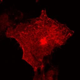 Immunocytochemistry/ Immunofluorescence - Anti-Lck antibody (ab3885)
Confocal immunofluorescence image of a COS cell transfected with a mouse Lck construct and stained with ab3885 at 1/200.
In untransfected cells, some nuclear staining is also visible as well as Lck staining.
Protocols
References (6)
ab3885 has been referenced in 6 publications.
- Lamm KYB et al. Inverted formin 2 regulates intracellular trafficking, placentation, and pregnancy outcome. Elife 7:N/A (2018). PubMed: 29309034
- Singh DK et al. SRC family kinases in hamster spermatozoa: evidence for the presence of LCK. Reproduction 153:655-669 (2017). PubMed: 28250239
- Luo T et al. Lck Inhibits Heat Shock Protein 65-Mediated Reverse Cholesterol Transport in T Cells. J Immunol 197:3861-3870 (2016). PubMed: 27742830
- Cao W et al. CXXC finger protein 1 is critical for T-cell intrathymic development through regulating H3K4 trimethylation. Nat Commun 7:11687 (2016). PubMed: 27210293
- Clayton KL et al. T cell Ig and mucin domain-containing protein 3 is recruited to the immune synapse, disrupts stable synapse formation, and associates with receptor phosphatases. J Immunol 192:782-91 (2014). WB . PubMed: 24337741
- Rüder C et al. The tumor-associated antigen EBAG9 negatively regulates the cytolytic capacity of mouse CD8+ T cells. J Clin Invest 119:2184-203 (2009). IHC (PFA fixed) ; Mouse . PubMed: 19620783
Images
-
Western blot - Anti-Lck antibody (ab3885)All lanes : Anti-Lck antibody (ab3885) at 1 µg/ml
Lane 1 : Jurkat (Human T cell lymphoblast-like cell line) Whole Cell Lysate
Lane 2 : Jurkat nuclear extract lysate (ab14844)
Lysates/proteins at 10 µg per lane.
Secondary
All lanes : Goat Anti-Rabbit IgG H&L (HRP) (ab97051) at 1/10000 dilution
Developed using the ECL technique.
Performed under reducing conditions.
Predicted band size: 57 kDa
Observed band size: 57 kDa
Exposure time: 4 minutes
-
Western blot - Anti-Lck antibody (ab3885)All lanes : Anti-Lck antibody (ab3885) at 1/1000 dilution
Lane 1 : COS cells transfected with mouse Fyn
Lane 2 : COS cells transfected with mouse Lck
Predicted band size: 57 kDaWestern blot against Lck using ab3885.
Lane 1: COS cells transfected with mouse Fyn.
Lane 2: COS cells transfected with mouse Lck.
-
Immunocytochemistry/ Immunofluorescence - Anti-Lck antibody (ab3885)
Confocal immunofluorescence image of a COS cell transfected with a mouse Lck construct and stained with ab3885 at 1/200.
In untransfected cells, some nuclear staining is also visible as well as Lck staining.


