Anti-JNK1+JNK2 (phospho T183 + Y185) antibody (ab4821)
Key features and details
- Rabbit polyclonal to JNK1+JNK2 (phospho T183 + Y185)
- Suitable for: ICC/IF, WB
- Reacts with: Mouse, Human
- Isotype: IgG
Overview
-
Product name
Anti-JNK1+JNK2 (phospho T183 + Y185) antibody
See all JNK1+JNK2 primary antibodies -
Description
Rabbit polyclonal to JNK1+JNK2 (phospho T183 + Y185) -
Host species
Rabbit -
Specificity
Phosphorylation site-specific antibody selective for the dually phosphorylated form of the c-Jun N-terminal Kinase (JNK)/Stress-Activated Protein Kinase (SAPK) enzymes containing a phosphate on threonine 183 and tyrosine 185 (human JNK 1 + 2). The antibody has been shown to recognize the endogenous, active forms of JNK 1 + 2 in a variety of cell types following treatment by a broad range of extracellular stimuli [e.g. including 293 cells (human embryonic kidney; +/- ultraviolet light) and PC12 cells (rat pheochromocytoma; +/- sorbital)]. The region of JNK1 and JNK2 surrounding T183 + Y185 has a high degree of similarity to the corresponding regions in JNK3 and thus may cross react with this protein if phosphorylated on the corresponding residues. -
Tested applications
Suitable for: ICC/IF, WBmore details -
Species reactivity
Reacts with: Mouse, Human
Predicted to work with: a wide range of other species
-
Immunogen
Synthetic peptide (Human) derived from the region of JNK 1 + 2 that contains threonine 183 and tyrosine 185, based on the human sequence. This region is conserved among many species including human, rat, mouse, chick, nematode (Caenorhabditis elegans), and fly (Drosophila melanogaster).
Properties
-
Form
Liquid -
Storage instructions
Shipped at 4°C. Upon delivery aliquot and store at -20°C or -80°C. Avoid repeated freeze / thaw cycles. -
Storage buffer
pH: 7.30
Preservative: 0.05% Sodium azide
Constituents: PBS, 50% Glycerol, 0.1% BSA
BSA is IgG and protease free -
 Concentration information loading...
 -
Purity
Immunogen affinity purified -
Purification notes
Purified from rabbit serum by sequential epitope specific chromatography. The antibody has been negatively preadsorbed using a non-phosphopeptide corresponding to the site of phosphorylation to remove antibody that is reactive with non-phosphorylated JNK enzymes. The final product is generated by affinity chromatography using a JNK-derived peptide that is phosphorylated at threonine 183 and tyrosine 185, within the activation loop. Note: It is the dually phosphorylated form of these enzymes that has full enzymatic activity. -
Clonality
Polyclonal -
Isotype
IgG -
Research areas
Images
-
MEF1 cells were incubated at 37°C for 48h with vehicle control (0 µM) and 5 µM of glibenclamide (ab120267) in DMSO. Increased expression of of JNK1+JNK2 (phospho T183 + Y185) (ab4821) correlates with an increase in glibenclamide concentration, as described in literature.
Whole cell lysates were prepared with RIPA buffer (containing protease inhibitors and sodium orthovanadate), 10µg of each were loaded on the gel and the WB was run under reducing conditions. After transfer the membrane was blocked for an hour using 3% milk before being incubated with ab4821 at 1/1000 dilution and ab85139 at 1 µg /ml overnight at 4°C. Antibody binding was detected using an anti-rabbit antibody conjugated to HRP (ab97051) at 1/10000 dilution and visualised using ECL development solution.
-
ab4821 staining JNK1 + JNK2 (phospho T183 + Y185) in A549 cells (green, panel a) by ICC/IF (Immunocytochemistry/immunofluorescence). Cells were fixed with 4% paraformaldehyde, permeabilized with 0.25% Triton X-100 and blocked with 5% BSA for 1 hour at room temperature. Samples were incubated with primary antibody (2ug/ml in 1% BSA) for 3 hours at room temperature. An Alexa Fluor® 488-conjugated Goat anti-rabbit IgG polyclonal was used as the secondary antibody (1/400). Nuclei stained with DAPI (blue, panel b), F-actin stained with Alexa Fluor® 594 Phalloidin (red, panel b) and merged images (panel d).
-
To demonstrate the phosphorylation of JNK 1 & 2 in a cell based assay, 293 cells were treated with ultraviolet irradiation (UV). Proteins from cell extracts were resolved by SDS-PAGE on a 10% Tris-glycine gel and transferred to nitrocellulose. Membranes were incubated with either 1
µ g/mL ab4821 or 1µ g/mL anti-JNK1 pan. After washing, membranes were incubated with goat F(ab’)2 anti-rabbit IgG alkaline phosphatase and bands were detected using the Tropix WesternStar detection method.
To demonstrate the phosphorylation of JNK 1 & 2 in a cell based assay, 293 cells were treated with ultraviolet irradiation (UV). Proteins from cell extracts were resolved by SDS-PAGE on a 10% Tris-glycine gel and transferred to nitrocellulose. Membranes were incubated with either 1 µ g/mL ab4821 or 1 µg/mL anti-JNK1 pan. After washing, membranes were incubated with goat F(ab’)2 anti-rabbit IgG alkaline phosphatase and bands were detected using the Tropix We -
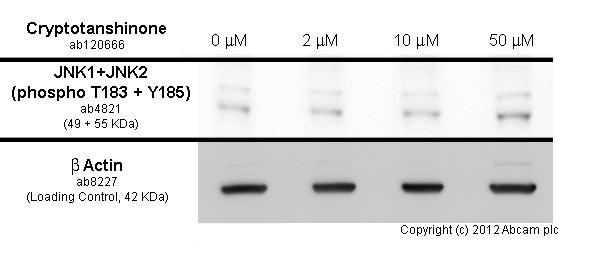
MCF7cells were incubated at 37°C for 4h with vehicle control (0 µM) and different concentrations of cryptotanshinone (ab120666). Increased expression of JNK1+JNK2 (phospho T183 + Y185) in MCF7 cells correlates with an increase in cryptotanshinone concentration, as described in literature.
Whole cell lysates were prepared with RIPA buffer (containing protease inhibitors and sodium orthovanadate), 10µg of each were loaded on the gel and the WB was run under reducing conditions. After transfer the membrane was blocked for an hour using 5% BSA before being incubated with ab4821 at 1/1000 dilution and ab8227 at 1 µg/ml overnight at 4°C. Antibody binding was detected using an anti-rabbit antibody conjugated to HRP (ab97051) at 1/10000 dilution and visualised using ECL development solution.
-
 Immunocytochemistry/ Immunofluorescence - Anti-JNK1+JNK2 (phospho T183 + Y185) antibody (ab4821) This image is courtesy of an Abreview submitted by Mr George Chennell
ab4821 staining JNK1+JNK2 (phospho T183 + Y185) in human foreskin fibroblasts by ICC/IF. The cells were fixed in cytoskeletal fixative, permeabilized in 0.5% Triton X-100 and blocked in 2% dillution buffer (2%BSA + 0.1% Triton X-100) for 1 hour at 25°C. The primary antibody was diluted, 1/100 and incubated with sample for 12 hours. An Alexa Fluor® 594 conjugated goat polyclonal to rabbit IgG, diluted 1/250 was used as secondary.


